The post Seek Shade and Use Sunscreen Daily to Shield Yourself from Harmful Rays appeared first on University Health News.
]]>Some other reasons sun protection may be even more of a priority during summer is that in many locations daylight hours are longer in the summer months; many of us engage in outdoor activities such as swimming, hiking, and picnicking; and women tend to wear lighter, more revealing clothing during the warmer months, exposing more skin to the sun. Regardless of the season, it’s essential to make sun protection part of your daily routine, including applying sunscreen, seeking shade, wearing protective clothing and sunglasses, and avoiding outdoor activities during peak sun hours when possible.
Minimize Skin Cancer Risk
Skin cancer is more prevalent in older adults due to cumulative sun exposure over their lifetime. About 90 percent of nonmelanoma skin cancers, which includes basal cell carcinoma (BCC) and squamous cell carcinoma (SCC), are associated with exposure to UV radiation from the sun, according to the Skin Cancer Foundation. UV exposure is also a major risk factor for the development of melanoma, which is less common than nonmelanoma skin cancers, but is more aggressive and has a higher risk of spreading to other parts of the body, according to the American Cancer Society.

Protecting your skin from the sun’s harmful rays can reduce the risk of developing skin cancer.
Studies show that regular sunscreen use can reduce the risk of BCC, the most common type of skin cancer, by up to 50 percent. “Both UVA and UVB rays contribute to skin cancer risk, so it is important to use a sunscreen that protects against both types of rays. Buying a product labelled “broad-spectrum” will ensure that you are protected against both types of rays,” says Shari Lipner, MD, PhD, associate professor of clinical dermatology at Weill Cornell Medicine. The American Academy of Dermatology (AAD) recommends using a broad-spectrum sunscreen with an SPF (sun protection factor) of 30 or higher.
Regardless of ethnicity or race, no one is immune from the sun’s damaging effects. While people with darker skin have more natural protection against UV radiation compared with those with lighter skin, they are still susceptible to sunburn, skin damage, and skin cancer. “To drive home the importance of using sunscreen to all of my patients, I tell them a story about one of my patients (without revealing the patient’s identity) who developed skin cancer and then had to face all of the consequences, including surgery, that went along with that,” says Dr. Lipner.
Protect Your Skin All Year
UVA and UVB rays are present all year long, in all climates and regions, even on cloudy or overcast days. These rays can penetrate clouds and reflect off surfaces such as snow, water, sand, and pavement, increasing the risk of sunburn, skin damage, and skin cancer. Sunscreen should be a part of everyone’s daily self-care routine. For women who use facial foundation, CeraVe’s Hydrating Mineral Sunscreen is a good combination product: It offers full-spectrum coverage, with an SPF of 30.
Here are some tips to follow for effective sunscreen use:
- All exposed skin should be covered with a layer of sunscreen. A shot glass, or one ounce, is generally enough to cover all the skin.
- Sunscreen should be reapplied every two hours and more often after sweating or swimming.
- Check the expiration date. If the sunscreen is expired, or there’s no expiration date but it’s more than three years old, throw it out and get a new product.
Prevent Premature Aging
In addition to skin cancer prevention, there are other reasons to protect your
skin from the sun. For example, prolonged sun exposure can lead to premature aging of the skin, including wrinkles, fine lines, age spots, and sagging skin. Sun exposure can also exacerbate conditions, such as eczema and psoriasis, and can cause uneven pigmentation leading to sunspots, freckles, and hyperpigmentation that can appear as brown, black, gray, or pink patches.
Schedule an Annual Skin Exam
Regular skin examinations by a health-care professional are important for early detection of skin cancer. By including broad-spectrum sunscreen in your daily skin-care routine and wearing protective clothing, hats, and sunglasses, you can protect yourself from the sun’s harmful rays and reduce the risk of sunburn, skin damage, and skin cancer.
The post Seek Shade and Use Sunscreen Daily to Shield Yourself from Harmful Rays appeared first on University Health News.
]]>The post Could That Spot Be Skin Cancer? appeared first on University Health News.
]]>Know Your Risk Factors Skin cancer typically develops on skin that has been exposed to the sun over a long period of time—hence, older age increases your risk. You’re also more vulnerable if you have a family history of skin cancer, and/or have already had skin cancer. Your skin type is a factor too, with fair-skinned people particularly susceptible, especially if they have red or blonde hair.
Other risk factors include burning easily with sun exposure; having sustained blistering sunburns in your youth; and having lots of freckles, moles, and/or actinic keratoses (scaly pink or brown patches that are considered to be the earliest stage in the development of skin cancer). But don’t assume that not having these risk factors means you’re safe from skin cancer. “While people with darker skin and hair are less likely to get skin cancer, they still can develop it,” confirms Dr. Akhavan.
Lastly, some medications commonly used by older adults—including nonsteroidal anti-inflammatory drugs (NSAIDs) like aspirin and ibuprofen (Advil®, Motrin®), some antibiotics, and diuretics can increase your skin’s sensitivity to the sun. Where the latter are concerned, research has suggested that people who take hydrochlorothiazide (HCTZ) may be significantly more likely to develop non-melanoma skin cancers than people who do not take HCTZ.

Protecting Your Skin The best protection against sun damage is sunscreen with a sun protection factor (SPF) of at least 15 and preferably 30. Dr. Akhavan recommends you wear it even on cloudy days, applying it about 30 minutes before going out to ensure that your skin absorbs it. “Reapply it every two hours or so, and if you sweat heavily or go swimming,” he adds. “Replace sunscreen as soon as it expires, since expired products won’t protect you as effectively.”
For creams or lotions, you should use about a palmful of sunscreen (you may need more, depending on how much skin is exposed). As well as the obvious areas (face, neck, shoulders, arms, legs), be sure to apply it to your scalp where you part your hair and all areas where hair is thinning (unless you’re wearing a sunhat, which remains the best option); eyelids; ears; lips (use a sunscreen stick); hands; and feet. A “baby” sunscreen that contains zinc oxide or titanium dioxide is less likely to irritate your eyes (you also may be less likely to get it in your eyes if you use a sunscreen stick). If using a spray, overlap each pass with the container to avoid missing any skin.
Ideally, you should try to keep out of the sun when it is at its strongest (between 10am and 4pm). If you do spend time outdoors during that time, wear a wide-brimmed hat to provide additional protection for your face and the back of your neck.
Checking Your Skin Schedule a yearly skin examination with a dermatologist (every six months if you’re high risk), and set 10 to 15 minutes aside each month to check your skin yourself, using the ABCDE formula (see What You Can Do). Perform your self-check in front of a full-length mirror in good light, and don’t forget your scalp, underneath your breasts, your finger and toenails, the soles of your feet, and in between your toes. Use a hand mirror to check your back and genitals. “Keep a log detailing the location and description of moles, large freckles, and other marks on your skin, and if you have a smartphone, take a picture,” Dr. Akhavan advises. “Having a record can help you better judge whether a lesion is new or an existing mark has changed.”
Stay Alert Finally, if you’ve already had skin cancer, keep in mind that it may raise your risk for other cancers, and stay up-to-date on routine screening tests, such as colonoscopy and mammogram. Also address other cancer risk factors by eating a healthy diet, limiting alcohol, getting plenty of exercise, and not smoking.

The post Could That Spot Be Skin Cancer? appeared first on University Health News.
]]>The post How to Tell the Difference Between Age Spots and Skin Cancer appeared first on University Health News.
]]>How to Identify Age Spots
If you’re 50 or older, you’re likely to notice new age spots developing on your hands, face, and other areas of your skin that have received the most sun exposure. “Solar lentigines” is the medical term for these areas of skin discoloration that are commonly called “age spots” or “liver spots.”
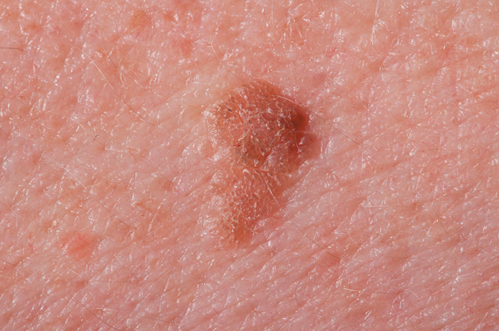
“Lentigines are sharply defined patches that are tan or light brown in color. Benign (non-cancerous) moles are typically brown to darker brown, and they may be flat or raised. Melanomas may be larger and darker than moles,” explains Shari Lipner, MD, PhD, a dermatologist at Weill Cornell Medicine. (See sidebar below for more guidance on identifying melanomas.)
Dr. Lipner says that lentigines may form due to a genetic disposition or as a result of sun exposure. Lentigines are not cancerous or dangerous, but people who have significant past sun exposure are more likely to have them, as well as an increased risk of developing skin cancer.
Signs of Skin Cancer: The Importance of Screening
Age spot or liver spot.
If you’re at risk of skin cancer, it’s wise to have a full-body exam by a dermatologist every one to two years. A melanoma may not look very threatening, but the longer it goes undetected and untreated, the more likely the cancer will spread to other areas of your body.
“A board-certified dermatologist is best equipped to diagnose skin cancers. In addition to rigorous training, we also have special lighting and devices called dermatoscopes that help differentiate between lentigines, moles, and skin cancers,” says Dr. Lipner.
Between visits to your dermatologist, do a monthly self-check of your skin to monitor your moles, brown spots, and freckles, as well as any new spots or growths that appear. Perform your self-exam under a bright light, using a full-length mirror as well as a hand mirror. And, make sure to check your lips, mouth, ears, scalp, under your breasts, fingernails, and toenails, between your fingers and toes, the soles of your feet, and your genitals.
Know Your Risk of Skin Cancer
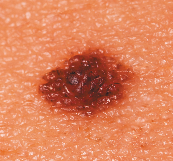
Non-cancerous mole.
One million people age 65 and older develop some type of skin cancer each year, and they also have the highest death rate from melanoma. Cumulative exposure to the sun is the main contributor to older adults’ higher risk.
You’re also at higher risk of skin cancer if you have:
- Fair skin
- Red or blond hair
- A history of blistering sunburns and/or outdoor summer jobs for three or more years as a teenager
- A large number of moles
- A history of actinic keratoses (scaly pink or brown patches that are considered to be precursors to skin cancer)
- Marked freckling of the upper back
- A personal or family history of the disease
Protect Yourself From Melanoma
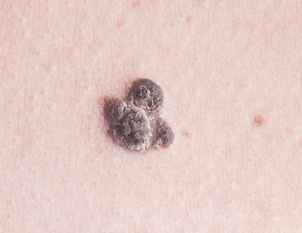
Melanoma.
Reducing your exposure to the sun’s UV rays is the only action you can take to lower your risk of skin cancer. (See also “On the Lookout for Basal Cell Carcinoma Symptoms.”)
If you spend time in the sun, even in the winter, use a sunscreen with a sun protection factor (SPF) of 30 or higher that is labeled “broad-spectrum;” this means that it protects against both UVA and UVB light. Use sunscreen even on cloudy days—about 80 percent of the sun’s rays can filter through clouds.
Apply sunscreen 15 to 30 minutes prior to sun exposure. Cover any exposed skin, including your ears, lips, back of your neck, and your scalp if your hair is thinning. For added protection, stay out of direct sunlight between 10 a.m. and 4 p.m.
SKIN CANCER WARNING SIGN: WHAT YOU CAN DO
Follow the “ABCDE” rule as a guide when examining moles and other spots on your skin. Look for:
- Asymmetry, in which one half of the mole is shaped differently than the other half.
- Borders that are irregular, ragged, or blurred.
- Color that varies from one area to another, with shades of tan, brown, black, white, pink, red, or blue.
- Diameter larger than 6 millimeters (the size of a pencil eraser).
- Evolving, such as growing larger and/or changing color or shape.
RECOMMENDED FOR YOU
- Signs of Melanoma Skin Cancer
- Battle of the Sexes: Skin Cancer Edition
- Performing Self-Exams Can Aid In Early Detection of Skin Cancer
The post How to Tell the Difference Between Age Spots and Skin Cancer appeared first on University Health News.
]]>The post Are You Protecting Vulnerable Areas From the Sun? appeared first on University Health News.
]]>Poor Application For the study, researchers looked at the efficacy of daily moisturizers that incorporate sun protection. “These moisturizers are widely available, but when they first became popular they tended to offer relatively low-grade protection against the sun,” says Arash Akhavan, MD, assistant clinical professor of dermatology at Mount Sinai. “More recently, these products frequently advertise a sun protection factor—or SPF—of 30 or greater, which most dermatologists recommend for optimum protection against the sun’s harmful ultraviolet rays.” However, as with sunscreens designed for use on the body, you won’t benefit from this protection if you don’t adequately cover the skin on your face.
One reason people don’t apply enough sunscreen on their face is that it can sting your eyes. The researchers hypothesized that this concern would be lessened among people using SPF-moisturizers, and that this would result in improved coverage. However, many of the 84 study participants consistently skipped applying the SPF-moisturizer to about 20 percent of the area around and on their eyelids. That’s a similar number to that seen among traditional sunscreen users in a previous study by the same research team.
Dr. Akhavan says the data are alarming given the disproportionately high incidence of melanoma (the most lethal form of skin cancer) and non-melanoma skin cancers—squamous cell carcinoma (SCC) and basal cell carcinoma (BCC)—on the head and neck compared with the rest of the body. “A 2018 study suggested that SCC of the eyelids is on the rise, while other research has indicated that the medial canthal area, which is the tiny patch of skin that connects the upper and lower eyelids at their inner corner, is particularly susceptible to BCC,” he observes. “While SCC and BCC are not as lethal as melanoma, they still can progress if not treated early.” Of course, prevention is better than cure, which is where sunscreen comes in. But previous research has shown that the medial canthal area is the least effectively covered region of the face when it comes to sunscreen application.
What Sunscreen Best Suits Your Eye Area? Dr. Akhavan says the best sun protection option for the skin around your eyes is an SPF-30 or greater sunscreen that contains zinc oxide or titanium dioxide, both of which are less likely to sting the eyes. “A sunscreen stick with these ingredients is ideal, since the stick formulation means the product is less likely to get into your eyes,” he says. “Apply the sunscreen all over your eyelids and under your eyes, right up to the eyelid margin, and don’t neglect the medial canthal area.” Also wear sunglasses with 100-percent ultraviolet protection. If you wear glasses, consider investing in transitional lenses that go dark when you’re out in the sun. Clip-on sunglasses are another option, but you also could opt for an inexpensive pair of large sunglasses that sit comfortably over your glasses—these may cover more of your eye area than clip-ons.
Protecting Other Vulnerable Areas Be sure to adequately protect your ears and lips, too. “Protecting the lips is especially important for people who take hydrochlorothiazide, which is used to lower high blood pressure,” Dr. Akhavan cautions. “The drug increases the skin’s sensitivity to the sun.” Other medications also may affect your skin in this way, so check drug labels and instructions for warnings about increased photosensitivity.
A wide-brimmed hat is advisable even if you are wearing sunscreen. Not only can it help protect your face and the back of your neck—it also ensures scalp protection, which is vital for people with thinning hair. “If you’re not fond of wearing hats, apply sunscreen to any areas of your scalp with sparse hair,” Dr. Akhavan adds.
It’s also important to use enough sunscreen. A quick dab or spray won’t cut it—you need about a shot glass full (or a palmful) of sunscreen if you’re using a cream or lotion (you may need more, depending on how much skin is exposed). “Apply the product 15 to 30 minutes before going out, to ensure that it is fully absorbed into your skin,” Dr. Akhavan advises.
Related post: “Medications to Treat Alzheimer’s Disease“
The post Are You Protecting Vulnerable Areas From the Sun? appeared first on University Health News.
]]>The post Dietary Fat Intake Linked to Skin Cancer Risk appeared first on University Health News.
]]>The post Dietary Fat Intake Linked to Skin Cancer Risk appeared first on University Health News.
]]>The post 8. Glossary appeared first on University Health News.
]]>allergic contact dermatitis: A rash that appears when the body’s immune system overreacts to foreign substances.
alpha-hydroxy acids: Acids derived from fruit and milk that are used in creams and lotions to try to reduce age spots, wrinkles, and other signs of aging.
antioxidants: Substances that protect cells from damage caused by unstable molecules known as free radicals, which are produced by the body as a normal byproduct of metabolism.
atopic dermatitis (atopic eczema): A skin disorder characterized by itching, scaling, and thickening of the skin that occurs in individuals predisposed to certain hypersensitive reactions.
basal cell carcinoma (BCC): The most common, most curable form of skin cancer.
basal cells: A cell in the deepest layer of the epithelium (the outside layer of cells that covers surfaces of the body).
biologic: A substance or product produced from one or more living organisms.
biomarker: A measurable substance in the body that indicates the presence or absence of disease or the risk for later developing a disease.
boil: A red, elevated, painful bump often caused by an infected hair follicle.
brachytherapy: Use of a machine to generate targeted doses of radiation administered through an applicator placed directly on the skin.
broad-spectrum sunscreen: One that protects against both types of ultraviolet radiation (UVA and UVB).
cellulitis: A common bacterial infection of the skin.
ceramides: Fatty molecules that limit the loss of water and act as a barrier against harmful substances.
chemotherapy: The use of drugs (taken either orally or intravenously) to kill cancer cells.
collagen: Fibers of protein found in connective tissue, cartilage, and bone.
contact dermatitis: A rash caused by something a person has come in contact with, such as a chemical or poisonous plant.
corn: A small patch of thickened, dead skin with a central core, usually caused by friction, and appearing between the toes or on the top or outer sides of the toes.
corticosteroids: Medications used to treat, among other conditions, some allergic skin conditions.
cryotherapy: Freezing an area of skin to destroy unwanted tissue, such as growths, moles, or warts.
curettage-electrodesiccation: Scraping off a cancerous area with a curette and burning any remaining cells with an electric current.
dermabrasion (surgical skin planing): A procedure that wears away the layers of skin and improves irregularities on the surface of the skin.
dermatologist: A physician who specializes in skin disorders.
dermis: The middle layer of skin that contains nerves, blood vessels, oil glands, and hair follicles.
dermatoscope: A handheld microscope-like device that magnifies a pigmented lesion and allows the dermatologist to see through the outermost layer of skin.
diabetic dermopathy: A skin disease specific to diabetes patients, characterized by brown, scaly, oval, or circular patches of skin on the front of the legs.
elastin: A protein in the skin that helps maintain resilience and elasticity.
emollients: Another name for moisturizers that soften the skin.
epidermal cyst: A sac beneath the surface of the skin filled with keratin and fatty material.
epidermis: The tough, outer protective layer of skin.
eruptive xanthomatosis: A skin disease that occurs in severe cases of diabetes, characterized by small bumps on the skin and surrounded by a red circle.
excisional surgery: A procedure in which a lesion or growth is surgically removed.
fitovers: Sunglasses that can be worn over prescription glasses.
folliculitis: A damaged hair follicle infected with staphylococcus bacteria.
granuloma: Small area of inflammation that can occur in many diseases.
herpes simplex: A virus that causes cold sores and blisters around the mouth.
hives: An allergic reaction of the skin characterized by red, raised marks in irregular sizes ranging from quite small to several inches.
humectants: glycerin and hyaluronic acid used to reduce the loss of moisture.
hyaluronic acid: A substance in connective tissues that cushions, lubricates, and provides volume to the skin.
immunotherapy: A type of treatment that boosts or restores the immune system to fight cancer, infections, and other diseases.
impetigo: A skin infection caused by bacteria and characterized by skin lesions that itch first and ooze later.
irritant contact dermatitis: A reaction that occurs when the skin is damaged by a foreign substance.
keratinocytes: Cells in the epidermis that produce keratin (a fibrous protein).
laser surgery: The use of high-intensity light to treat several diseases, including basal cell skin cancer.
lichen simplex: An itchy patch of skin caused by any one of several skin conditions.
lupus: A chronic disease in which the body’s immune system attacks its own cells, tissues, and organs.
melanin: The pigment that gives skin, hair, and eyes their color.
melanocytes: Cells in the basal layer of the epidermis that produce melanin.
melanoma: A malignant tumor associated with the most serious type of skin cancer.
Merkel cell carcinoma: A rare and aggressive form of skin cancer that appears as a hard patch or firm bump.
methicillin-resistant staphylococcus aureus (MRSA): A potentially dangerous staph bacteria resistant to certain antibiotics.
Mohs micrographic surgery: A procedure in which skin is removed layer by layer and then examined for the presence of cancer cells.
mole (nevus): A spot on the skin that contains melanin cells, which give color to the skin.
nevi: Birthmarks or moles.
nodular basal cell carcinoma: The most common BCC subtype.
nonsteroidal anti-inflammatory drug (NSAID): A substance that reduces inflammation and pain; ibuprofen, naproxen, and aspirin are examples.
occlusives: Petrolatum and other oily substances that block the evaporation of water.
photoaging: Damage caused by sun exposure over a period of time.
photodynamic therapy: The use of natural or UV light to treat psoriasis and other conditions.
phytonutirents: Substances found in certain plants believed to be beneficial for various health conditions, including skin.
psoriasis: A chronic, inflammatory, immune-related condition of the skin.
psoriatic arthritis: A form of arthritis that occurs in 10 to 30 percent of people who have psoriasis.
RDA: Recommendation Daily Allowance (of nutrients).
radiation therapy: The use of high-energy x-rays, particles, or radioactive seeds to kill cancer cells.
retinol: A compound made from vitamin A used to treat acne, aging skin, psoriasis, and certain skin cancers.
rosacea: A skin condition characterized by red skin that resembles a sunburn.
scabies: An infestation of the skin with a microscopic mite; more likely to affect older adults with weakened immune systems.
seborrheic keratosis: A benign skin tumor that is common in older adults.
shingles: A disease caused by the herpes zoster virus and characterized by pain and a skin rash.
skin tag: A benign, skin-colored growth on the skin; more common after age 60.
squamous cell carcinoma (SCC): The second-most common form of skin cancer.
squamous cells: Cells found in the outer layer of skin (epidermis).
subcutaneous layer (subcutis): The deepest layer of skin.
sun protection factor (SPF): A measurement of a sunscreen’s effectiveness, e.g., SPF 15, equating to how long a person wearing sunscreen can stay in the sun before beginning to burn.
T cells: White blood cells that circulate in the blood and lymph to activate the immune system’s response to infected or malignant cells.
ultraviolet protection factor (UPF): A measurement of how much ultraviolet radiation a particular clothing material absorbs.
ultraviolet A (UVA) rays: Rays that penetrate the skin deeply and are associated with wrinkling and sagging.
ultraviolet B (UVB) rays: Rays primarily responsible for sunburn.
urushiol: The toxic substance found in poison ivy, poison oak, and poison sumac.
vitiligo: A condition in which the skin or hair lose color (pigment).
wart: A noncancerous growth on the skin caused by a viral infection.
The post 8. Glossary appeared first on University Health News.
]]>The post 5. Skin Conditions: Lyme Disease to Wrinkles appeared first on University Health News.
]]>Many skin conditions involve some kind of lesion. A lesion simply means an abnormal growth or change in tissue, though the term is often associated with cancer. Most noncancerous skin lesions do not spread to other parts of the body, but benign lesions should nevertheless be self-monitored. If changes occur, have the lesion examined by a clinician. Look for new growth, moles that are increasing in size or color, and any growth that causes unusual itching, pain, or bleeding.
Some common skin growths may simply be unsightly (skin tags, for example), and many older adults prefer to seek medical attention to remove them. U.S. Pharmacist reminds us that because removal of benign skin growths are considered cosmetic, they may not be covered by Medicare or other health insurance plans. This means you have to decide between living with a skin tag or paying to have it removed.
Lyme Disease
Lyme disease is caused by four kinds of bacteria and spread when people are bitten by a blacklegged deer tick. But not all  ticks carry the bacteria and not all people bitten by the tick get the disease.
A characteristic feature of Lyme disease, and the key feature of early localized infection, is a slowly expanding red rash on the skin (called erythema migrans) at the site of the tick bite. The rash is often bull’s-eye shaped.
Poor sleep has emerged as a symptom of Lyme disease patients. A 2018 study published in the journal SLEEP found that people who have the condition report poor sleep quality before treatment and significantly worse sleep and sleep disturbance six months after treatment.
Contact your physician if you get a bulls-eye looking rash, flu-like symptoms, fever or chills, muscle or joint pain, headaches, fatigue, or weakness. Lyme disease is diagnosed by a physical exam and blood tests. If untreated, the disease can spread to the heart, joints, and nervous system.
Treatment with antibiotics is normally successful within a few weeks. If Lyme disease is detected later, antibiotics are still the treatment but it may take longer to get rid of the symptoms.
A person who has had Lyme disease once can get it again. There is no known vaccine, so reducing your exposure is the best way to prevent it.
Measles
 In June 2019, the highest number of measles cases in the United States (1,123) was reported by the Centers for Disease Control and Prevention (CDC) since 1992. The condition had been reported in 23 states.
The resurgence is due to misinformation about vaccines, the decline in vaccinated children, and foreign travel. Unvaccinated U.S. residents get the measles while they are abroad, bring the disease into the United States, and spread it to others.
The best way to protect against the disease is to get the measles-mumps-rubella shot (MMR). These three vaccines were combined in 1971. Rubella is also known as the German measles. The mumps is a contagious viral disease that attacks the salivary glands. Like the measles, mumps and rubella were common decades ago, but became rare due to the bundled vaccination program. If you were born prior to 1957, you were likely exposed to measles and have an adequate immune response. The CDC recommends that health-care workers, adults traveling overseas, college students, and anyone living near a community experiencing an outbreak get an MMR booster shot if they are unsure of their immunity. Another option is to get an inexpensive blood test (titer test), which can show if a you have enough measles antibodies to be protected against the disease. Claims that the MMR vaccine causes conditions such as autism and chronic diseases have long been scientifically discredited.
The CDC describes measles as a highly contagious viral respiratory illness that spreads through coughing and sneezing. It starts with a fever as high as 105 degrees Fahrenheit (°F), runny nose, cough, red eyes, and sore throat, and is followed by a rash that spreads over the body.
The rash usually appears about two weeks after exposure, and spreads from the head to the trunk and lower extremities. Those who get measles are contagious from four days before to four days after the rash appears. Some patients with compromised immune systems do not develop the rash. Those at high risk for complications are infants and children, adults over the age of 20, pregnant women, and those with impaired immune systems.
Very Contagious
Measles is one of the most contagious of all infectious diseases, warns the CDC. As many as nine of 10 susceptible persons  with close contact involving a measles patient will develop the disease. It can also be transmitted by breathing, coughing, and sneezing, and can remain in the air up to two hours after an infected person leaves the area.
There is no specific treatment, but symptoms could be addressed with acetaminophen (Tylenol) to reduce fever and relieve muscle pain, rest, and adequate hydration. The use of a humidifier might ease the symptoms of a cough and sore throat.
Moles
Moles are clusters or spots on the skin that consist of melanin cells called melanocytes, which give color to skin. They are either dark brown, reddish-brown, blue, or the color of skin. They vary in shape and size, but most are oval or round, and are less than one-quarter-inch in diameter.
Moles can be flat, raised, smooth, or wrinkled. Atypical moles are larger than a pencil eraser and usually have a dark brown center with a lighter, uneven border. They tend to run in families.
Bright red moles (cherry angiomas) are most likely to develop on the torso, arms, legs, or shoulders. They may begin to appear in a person’s 30s, and the number of moles tends to increase with age. They can be flat or slightly raised, round or oval-shaped, and range in size from a hardly-noticeable pinpoint to a quarter of an inch in diameter. Their reddish color is due to an abundance of small blood vessels. Red moles usually require no treatment unless you want them removed for cosmetic reasons or because they are bumped often and bleed. If you notice a change in the way a red mole looks, have a dermatologist examine it. If it has to be removed, it’s a relatively simple procedure of freezing it (cryosurgery), removing it by laser surgery, shaving it, or burning with an electric current (electrocauterization).
Most people have between 10 and 40 moles on average, and their presence was probably determined at birth. They can develop on any area of the body, including the scalp, between fingers or toes, and under fingernails and toenails.
The number of moles can change with age, with some fading as a person moves into adulthood, and others disappearing altogether or lasting 50 years or longer. Sunlight can increase the number of moles and make them darker.
Most moles are harmless and will never be a health threat, but in some cases they may lead to a higher risk of certain cancers. Traditionally, people who had the greatest number of moles were thought to have a greater risk of developing a melanoma.
A study in JAMA Dermatology suggested that physicians should not rely on the total number of moles as the only reason to perform an exam or determine a patient’s risk.
When to Call Your Doctor
The National Cancer Institute says to tell your doctor if you notice any of the following changes in a common mole:
- The color changes.
- The mole gets unevenly smaller or bigger.
- The mole changes in shape, texture, or height.
- The skin on the surface becomes dry or scaly.
- The mole becomes hard or feels lumpy.
- It starts to itch.
- It bleeds or oozes.
Moles or Melanomas?
University of Pennsylvania scientists appear to have discovered a gene (p15) that differentiates moles from melanomas.  By staining tissue samples, a low level of gene p15 indicated a melanoma. A high expression of p15 would be consistent with a benign mole.
A biopsy can determine if a mole is malignant, and a mole can be removed by shave excision (using a small blade to cut it out), punch biopsy (removing the mole with a small device that works like a cookie cutter), and excisional surgery (which involves taking out the mole and the skin around it). All of the procedures can be performed in a dermatologist’s office. Once removed, most moles do not recur. If one does, let your doctor know.
MRSA
Methicillin-resistant Staphylococcus aureus (MRSA) is a potentially dangerous staph bacterium that is resistant to certain antibiotics. It may cause skin and other infections throughout the body. The bacterium is carried by 2 percent of the population, but few are infected. Those who have weak immune systems are most vulnerable, especially those in hospitals, nursing homes, and other health-care facilities. The symptoms of MRSA may include a fever, and a bump or infected area of the skin that has one or more of the following conditions:
- Red, swollen
- Painful, warm
- Filled with pus
Draining and Medication
Treatment may involve draining the infection and taking an antibiotic, and it is important to continue taking the medication even if the infection appears to get better. Oritavancin (Orbactiv) is given in a single dose and may become an option for the treatment of MRSA, and other skin infections.
Researchers at the Los Angeles Biomedical Research Institute have challenged the current CDC strategy for dealing with MRSA in hospitals, which stresses limited contact between patients and staff. Their research showed that bathing patients in a common hospital soap called chlorhexidine was just as effective as limiting contact, and could result in better quality of care because of increased contact.
Three anti-MRSA antibiotics—dalbavancin (Dalvance), oritavancin, and tedizolid—have been approved by the U.S. Food and Drug Administration (FDA).
Preventing MRSA
The best ways to prevent MRSA are:
- Know the signs.
- Get treatment early.
- Practice good hygiene, especially by washing hands often.
- Do not share personal items, such as towels and razors.
- Follow prescription drug instructions precisely and complete the prescribed course, even if symptoms subside.
Take Your Medicine
A study published in Antimicrobial Agents and Chemotherapy found that patients with certain skin infections took, on average, only 57 percent of their prescribed antibiotic doses after leaving the hospital. Nearly half of 87 patients developed a new infection or needed additional treatment for the existing skin condition.
Poisonous Plants: Watch Out
Unless you live in Alaska, Hawaii, a desert, or at a high altitude, poison ivy is creeping around nearby. If you don’t stay away from it, you may be one of the 50 million Americans affected
each year.
Poison ivy, poison oak, and poison sumac all contain an oil called urushiol. It can cause an allergic reaction in the form of a blistering rash and an impossible-to-ignore itch. The poisonous plant presents three problems: recognizing it, treating it, and avoiding it.
Basic Summer Appearance
Poison ivy’s basic summer appearance is a vine or shrub that has green, three-leaf, pointed clusters. It can appear as ground cover, upright in bushes or shrubs, or as vines that grow up trees or rock walls. Several other plants (box elder, raspberries, blackberries) have a similar appearance. Poison ivy’s color changes with the seasons:
- In spring, the leaves emerge with a reddish color.
- In summer, they’re green. The plant may also have clusters of light green or cream-colored berries.
- In the fall, the leaves change to shades of red, orange, or yellow.
- In winter, its leaves disappear, but the leafless vines can still produce enough of the oil to cause a rash after contact.
Red Rash, Blisters, Itching
Poison ivy, oak and sumac are a year-round nuisance, but they are especially toxic in spring and summer. A nick or quick brush against the plant is all it takes. Its symptoms—a red rash, blisters, and itching—can appear within a few hours of exposure or as long as 12 days afterward. Without treatment, the symptoms go away in two to
three weeks.
If you know you’ve come into contact with poison ivy, oak or sumac flush your skin with lukewarm, soapy water as soon as possible (every minute counts), says the American Academy of Dermatology (AAD). Then do it again. Don’t scratch, and don’t break the blisters when they appear.
Oral antihistamines can relieve itching, as can wet compresses and short baths in warm or cool water. Over-the-counter topical corticosteroid preparations might help, or you might try trade-name products such as Calamine Lotion or Aveeno. See a doctor or go to the emergency room if:
- You have a temperature over 100°F.
- The affected area develops pus or soft yellow scabs.
- The rash spreads to your eyes, mouth, or genital area.
- You have trouble breathing.
- Home remedies haven’t done much to ease symptoms.
Not Contagious
There is some good news regarding a mostly-bad-news situation. The rash is not contagious and does not spread. It might appear to be spreading, but it’s because of a delayed reaction to urushiol in the areas of skin affected. Prevent exposure with these strategies:
- Wear long sleeves and pants tucked into garden boots if you are going to be working near poison ivy, oak or sumac. Use gloves that don’t allow fluids, including oils, to pass through.
- Wash any item of clothing that has been in contact with poison ivy, oak or sumac, and regularly wash garden tools.
- Use pet shampoo to wash a dog that may have brushed against one of these plants. Pets aren’t sensitive to the oil, but if it gets on their fur and you touch it, you’ll have a reaction.
- Don’t put poison ivy, oak or sumac leaves in a campfire or when burning leaves in your yard. Urushiol can become airborne.
Protection by Avoidance
Poison ivy, oak, and sumac can be easily pulled out in early spring if only a few plants are involved. Pull out the entire root system, if possible, and put it in a plastic bag to be taken away.
When the plants have been in an area for a long time, it is almost impossible to remove the plant and its root system. The best way to protect yourself is to stay away from it.
Plaque Psoriasis
 Even though psoriasis affects up to 8 million people in the United States, about 40 percent of psoriasis patients do not get treatment. There are five types of psoriasis, and each has unique symptoms, but 80 percent of people get the most common variety: plaque psoriasis.
Risk Factors
Psoriasis and other diseases are associated with metabolic syndrome (a group of risk factors associated with diabetes and stroke that include a large waistline, high triglycerides, low HDL cholesterol, high blood pressure, and high fasting blood sugar). Psoriasis may be a precursor to diabetes and cardiovascular disease. Approximately one-third of psoriasis patients have a family history of the disease.
T-Cells Rising
Psoriasis occurs when T-cells that normally protect the body against infection and disease develop and rise to the surface at a faster-than- normal rate. They accumulate on the top layer of skin before they have time to mature.
The discovery of a mutation in the CARD14 gene suggests that the mutation, plus an environmental trigger, is enough to cause psoriasis. Scientists now have a much clearer picture of what is happening in psoriasis, and targeted therapies are being developed.
Skin cell turnover usually takes about a month, but in psoriasis it can happen in a few days. This process results in patches of thick, inflamed skin covered with scales that itch and can hurt.
They appear anywhere on the body, but show up  most often on the elbows, knees, legs, lower back, face, palms, soles of the feet, and scalp. The symptoms can be only a nuisance or serious enough to interfere with work, recreation, and daily life and functions.
Patches, Scales, Cracked Skin
Symptoms include red patches, silver scales, dry skin, cracked skin that can bleed, thick or ridged nails, and swollen or stiff joints. A new episode can be triggered by infections, injuries to the skin, smoking, cold temperatures, stress, alcohol consumption, and certain medications, including lithium, beta blockers, and antimalarial drugs. Excessive body weight also can increase the risk of psoriasis.
Excessive inflammation is a critical feature, and since it is also a characteristic of insulin resistance, obesity, high cholesterol levels, and cardiovascular disease, psoriasis patients should consult with their health-care providers to watch closely for signs of these conditions.
Treatable, Not Curable
Psoriasis can be difficult to diagnose. It is not curable, but it is treatable. See a doctor if your condition is more than a nuisance, if it interferes with daily activities, and if you are concerned about the appearance of your skin. If your doctor cannot make a diagnosis by observation, he or she will take a skin sample and examine it under a microscope.
Physicians often use a three-step treatment approach, which includes topical medication, phototherapy, and oral or injectable drugs.
Step 1 (one or more of the following):
- Corticosteroids to reduce inflammation and slow cell turnover.
- Calcipotriene, a synthetic form of vitamin D, to limit cell production.
- Retinoids, a synthetic form of vitamin A, to normalize DNA activities in cells.
- Moisturizers to reduce itching, scaling, and drying.
Step 2 (to calm cellular response):
- Photodynamic therapy to slow the rate of cell turnover.
Step 3 (one or more of the following):
- Methotrexate (Rheumatrex, Trexall)
- Cyclosporine (Gengraf, Neoral)
- 6-Thioguanine
- Hydroxyurea (Droxia, Hydrea, Siklos)
- Antibiotics
- Biologics
At-Home Treatment
The AAD suggests several strategies for dealing with psoriasis. They include:
- Bathe daily to remove scales and reduce inflammation, but avoid hot water and harsh soaps.
- Use moisturizers to prevent drying of the skin.
- Cover affected areas at night with moisturizers, and lightly cover with plastic food wrap.
- Use cortisone to reduce inflammation, such as over-the-counter creams with 0.5 to 1 percent cortisone.
- Avoid triggers such as stress, smoking, excessive sun exposure, and drinking alcohol.
- Try not to scratch.
Alternative Therapies
Although they do not always report it to their physicians, significant numbers of people are using alternative methods to treat skin conditions. In the case of psoriasis, a recent study found that up to 51 percent of patients with psoriasis were using complementary or alternative medical approaches (see “Many Psoriasis Patients Use Alternative Therapies”).
Psoriatic Arthritis
Psoriatic arthritis is a form of inflammatory arthritis. Its symptoms vary widely and make it difficult to diagnose, even for physicians. X-rays, joint fluid tests, and blood  tests are used to make the diagnosis.
Patients report pain and stiffness in the knees, ankles, and joints of the feet, although other areas can be affected. Inflamed joints may be swollen and hot, and stiffness and pain are often worse in the morning than at other times. Patients also may have inflamed tendons and inflammation in the eyes, lungs, and aorta. Acne is a common side effect.
Psoriatic arthritis can develop at any age, but it most often occurs in adults between ages 25 and 50. Genes, environmental factors, and the immune system appear to play a role in its development. Psoriasis precedes psoriatic arthritis by months or even years.
Treat the Symptoms
The goal of treatment is to suppress symptoms rather than cure the disease. Doctors first try nonsteroidal anti-inflammatory drugs, such as aspirin, ibuprofen (Advil, Motrin), and naproxen (Aleve, Naprosyn).
If they don’t work, disease-modifying antirheumatic drugs, such as methotrexate, antimalarials, sulfasalazine, cyclosporine, and tumor necrosis factor (TNF) inhibitors, are prescribed. TNF drugs, such as etanercept (Enbrel), infliximab (Remicade), and adalimumab (Humira), are effective for psoriasis and psoriatic arthritis.
The FDA has approved the drugs golimumab (Simponi) and ustekinumab (Stelara) for the treatment of psoriatic arthritis. They also have approved a drug called apremilast (Otezla) to treat psoriatic arthritis by blocking enzyme-related inflammation.
A rheumatologist should treat psoriatic arthritis if arthritis is the focus. If the primary symptoms involve only the skin, a dermatologist can treat them.
Rocky Mountain Spotted Fever
Don’t let the name fool you. Although the Rocky Mountain states are where it was first identified, Rocky Mountain spotted fever is more common in the southeastern and southcentral states than in other parts of the country.
Rocky Mountain spotted fever is caused when an infected wood tick or yellow dog tick bites a person. People are bitten most often from March to September, when the ticks are active and people are in tick-infested areas.
Rash, Headache, Fever
Rocky Mountain spotted fever is a serious and sometimes deadly bacterial infection that begins with a rash, headache, and high fever that can get worse quickly if it’s not diagnosed and treated. The rash does not itch, but it spreads rapidly from the wrists and ankles in both directions to the palms, soles, forearms, necks, armpits, buttocks, and trunk—however, 10 percent of people with the condition do not develop a rash.
Untreated cases of the disease result in death up to 80 percent of the time. Early treatment with antibiotics has reduced the death rate to about 5 percent.
Prevention
- Tuck trousers into shoes, socks, or boots.
- Stay on paths and trails.
- Wear light-colored clothing to spot ticks more easily.
- Wear long-sleeved shirts.
- Use tick repellants containing DEET.
- Search quickly for tick bites.
- Remove ticks immediately.
Rosacea
Rosacea is a common, chronic, incurable condition characterized by redness and visible blood vessels in the face. It has been classified into four subtypes, which are  described by the National Rosacea Society (NRS). See “Rosacea Subtypes and Symptoms.” More than 16 million people in the United States have rosacea and most of them don’t know it.
Cheeks, Chin, Nose
The cheeks, forehead, chin, and nose are the primary targets of rosacea. The neck, ears, chest, and back are less frequently involved. In 50 percent of people with rosacea, the eyes may be watery, red, or irritated. Other possible symptoms are a sensation of burning, itching, or stinging, dry or thick skin, and facial swelling.
Individuals most susceptible to rosacea have fair skin, blush easily, are between ages 30 and 60, and have a family history of the condition. Women are affected more often than men, but that may be because men wait longer to seek treatment.
Cause Unknown
The cause of rosacea is unknown, but recent research revealed that an immune response might play a role in its development. Through skin biopsies, a team of international researchers found that people with rosacea had unusually high levels of cathelicidins (types of proteins), peptides, and inflammatory properties that protect skin against infection. They also discovered that an enzyme known as SCTE was elevated in people with rosacea.
Family History
A family history of rosacea places people in a higher risk category, as do high rates of blistering sunburn. The NRS reports a connection between rosacea and national origin. Individuals with Irish, English, and German ancestry appear to be at higher risk.
Psychological Consequences
Rosacea has psychological as well as physical consequences. A survey conducted by the NRS found that more than 75 percent of patients said their condition lowered their self-confidence and self-esteem. Fifty-two percent said it was the reason they avoided public contact and frequently cancelled social engagements.
Risk of Cancer Increased
A study of 49,475 rosacea patients in Denmark found that, when compared to a control group of more than 4 million people, those with the condition had higher rates of liver cancer, non-melanoma skin cancer, and breast cancer, but a decreased risk of lung cancer. The findings were published in the journal Cancer Epidemiology.
If rosacea is not treated, it always gets worse, and sometimes progresses into a more serious condition called rhinophyma, which affects the nose, giving it a red, bulbous appearance. Ninety percent of rosacea patients say their condition is controlled with treatment.
Medications Take Time
Topical medications include azelaic acid, benzoyl peroxide, clindamycin, erythromycin, metronidazole, sulfacetamide, and sulfur lotions. Keep in mind that it might take two or three months to get significant results. In 2014, the AAD issued a statement acknowledging that skin prone to acne or rosacea may improve with daily probiotic use.
Studies specifically associating probiotics and rosacea are scarce. A 2017 study in Dermatology Practical & Conceptual concluded, “[Rosacea] patients may be advised to support a healthy gut microbiome, including the consumption of a fiber-rich [prebiotic] diet.” Given that proper microbe balance within the gut is beneficial, research continues on how probiotics might help reduce the inflammatory responses that result in acne and rosacea.
Pimples and bumps may respond better to oral antibiotics, including doxycycline (Adoxa, Monodox), erythromycin, minocycline, and tetracycline (Periostat, Vibramycin).
Oral antibiotics also may be combined with glycolic acid peels and glycolic washes and creams. Isotretinoin is not approved by the FDA for this condition, but some doctors prescribe it (off-label use) to help shrink facial skin that has thickened. However, it can have serious side effects, such as nosebleeds, dry skin, dry mouth, and itching.
The FDA approved the topical cream ivermectin (Soolantra) in 2014 for the treatment of inflammatory lesions of rosacea. Subjects who used the cream achieved “clear” or “almost clear” rates 38 to 40 percent of the time, compared with 7 and 19 percent of the time in a control group.
When the eyes are affected, patients should gently clean their eyelids with diluted baby shampoo, or an over-the-counter eyelid cleaning substance. Warm compresses applied several times a day also might relieve the symptoms. Oral antibiotics, such as doxycycline, minocycline, or tetracycline, are prescribed at times.
Dermatologists may use electrosurgery or laser surgery to address redness and flushing. Among recent innovations are pulsed dye lasers that destroy visible blood vessels and reduce flushing and redness. Intense pulsed light therapy delivers light to the affected areas, where it targets blood vessels and redness.
Avoid Triggers
 Rosacea cannot be prevented, but the symptoms can be controlled once they have developed. The first step is to avoid possible triggers. “Rosacea Triggers by Percent Affected” shows the results of a survey taken by the NRS of more than 1,000 patients regarding factors that produce an episode.
Use a 30 to 50 SPF sunscreen when outdoors, and a moisturizer during winter to prevent dry skin. Consider keeping a diary to identify substances, activities, and environments that could possibly cause a flare-up.
Scabies
Older adults, especially those with weakened immune systems, are at a higher risk for scabies than younger, healthier people. Scabies is an infestation of the skin by a microscopic mite. It is a common condition found around the world, and spreads rapidly in crowded conditions where there is frequent skin-to-skin contact. It can happen in hospitals, nursing homes, and other institutions, but prolonged contact is usually needed to transmit the infection. A casual handshake or hug is unlikely to cause a problem.
It could take four to six weeks after contact for symptoms to develop. The symptoms include skin irritation or a rash, usually between the fingers or on the wrists, elbows, knees, breasts, or shoulder blades. Your doctor can prescribe a topical cream or lotion to get rid of the infestation, although the symptoms may last a week or two longer.
Seborrheic Keratosis
Seborrheic keratosis (SK) looks like a wart-like tumor on the surface of the skin and may be mistaken for skin cancer. It is neither, but SK is a common skin growth in older adults.
SK can be yellow, brown, black, or other colors. The lesions are more common on the face, chest, shoulders, and back than on other places of the body. Some of them turn black, which is why they may be mistaken for skin cancer, but biopsies almost always determine them to be noncancerous.
SK can appear alone or in clusters, have a rough or smooth texture, and do not penetrate deeply into the skin. They can be quite small or more than one inch in diameter and are painless. However, rubbing or scratching can cause inflammation.
Family History
Descriptions of SK include the term “stuck on” or “pasted on,” as if someone dabbed a patch of candle wax onto the skin. They are not contagious, but they do seem to run in families.
Their cause is unknown, though as the name suggests, SKs emit an oily, waxy substance similar to that produced by sebaceous glands. Expert opinions are mixed as to whether ultraviolet (UV) light exposure causes SK.
The AAD says exposure to sunlight does not seem to be a cause or a complicating factor after SK has developed. However, others say exposure to sunlight may be a factor because the condition is common in areas often exposed to the sun.
Unsightly, Not Dangerous
The best news about SK is that it is not dangerous, and usually does not need to be removed. People who have them removed do so because of their unsightly appearance or because the growths get irritated, itch, or bleed. Insurance companies do not cover procedures to remove them for cosmetic purposes.
Also, do not waste your money on creams, ointments, or other over-the-counter products that claim to remove the growths. They don’t work.
A dermatologist can remove an SK during an office visit by cryosurgery (using liquid nitrogen), curettage (scraping them off the skin), electrosurgery (by means of an electrical current, sometimes combined with curettage), or laser surgery (using high-intensity light beams to destroy the growth). In 2017, the FDA approved ESKATA, a topical solution for the treatment of seborrheic keratosis.
Don’t confuse seborrheic keratosis with actinic keratosis. Actinic keratosis, also called solar keratosis, is considered to be the earliest stage of skin cancer that is limited to the outermost layer of skin.
Shingles
Almost one in three people in the United States will develop shingles during their lifetime. Two-thirds get it after the age of 50. Most will get it once, but it’s possible to get it a second or third time.
Shingles causes a rash or blisters on the skin. It is a viral disease—the same one that causes chickenpox. After chicken pox heals, the virus becomes dormant in the body, but it can emerge again if a person’s natural resistance is compromised.
Those who are at the greatest risk for shingles have a weakened immune system, are over the age of 50, have been ill, are experiencing trauma, and/or are under stress.
The symptoms of shingles often include itching, stabbing, or shooting pains. The skin appears red in the affected area. Other symptoms are fever, chills, headache, and an upset stomach. A rash appears after a few days around the waistline or on one side of the face or trunk.
Antiviral Medications
There is no cure for shingles, but treatment with antiviral medications—such as acyclovir (Zovirax), valacyclovir (Valtrex), and famciclovir (Famvir)—can help ease the pain and discomfort and reduce the duration of symptoms.
The vaccine Shingrix was approved by the FDA in 2017. The CDC recommends that healthy adults 50 years and older get two doses of Shingrix, two to six months apart. Pain, redness, swelling at the injection site, and muscle pain are possible side effects.
A review of 27 studies involving more than 2 million patients found that Shingrix was 97 percent effective in preventing shingles compared to Zostavax, which was 51 percent effective. (see “Shingrix More Effective Than Zostavax”).
Skin Tags
A skin tag is a small, benign, elongated, skin-colored growth that is common in people older than age 60. The condition is also known as an acrochordon. It is made up of a core of fibers and ducts, nerve cells, fat cells, and a covering. Some have a stalk.
 A tag sticks out of or looks like it is hanging from the skin, usually near the neck, armpits, trunk, breasts, or other areas of the body where the skin folds. Some may be darker than the skin color.
Skin tags occur in approximately 46 percent of the population, and the incidence increases with age. The number goes up to 59 percent by the age of 70.
Skin tags are present in both men and women, although they are associated with pregnancy in some women. Skin tags are also more common in people with type 2 diabetes and in those who are obese. Two studies have found that people who have multiple tags were more likely to have insulin resistance.
Skin tags appear to have an association with metabolic syndrome. There is some evidence that susceptibility may be genetic.
Skin tags do not grow (they are usually just a fraction of an inch in diameter), they do not hurt, and they are not a form of skin cancer. The can be unattractive and become irritated if clothes or jewelry rub against them. They may develop because of skin rubbing against skin and are more common in those who are overweight or have diabetes. Unusually large tags may burst and bleed under pressure.
Quick, Painless Treatment
A skin tag is easily diagnosed and treated. It can be removed during an office visit by excision (cutting it out), cryotherapy (freezing it), cauterization (burning it off with an electrical current), or ligation (interrupting the blood supply). Removing larger tags may require a local anesthetic. A skin tag on the eyelid may have to be removed by an ophthalmologist.
The National Institutes of Health (NIH) do not recommend that a person attempt to remove a skin tag without medical assistance because of the risk of bleeding and infection. Over-the-counter products are available, but not recommended by any national health organization in the United States.
Tags do not normally come back at the same site, but new growths can develop elsewhere on the body. There is no evidence that removing a skin tag causes more to develop. Some tags just fall off, but in most cases, they don’t.
You cannot prevent skin tags—they just happen. But unless they regularly become irritated, unsightly, or change in color, size, composition, or sensitivity, there is no reason to treat or report them.
Sunburn Consequences
Many skin diseases are caused by overexposure to the sun. If the sun does not cause them, many are made worse by exposure to the sun’s UV rays. However, there are benefits to sun exposure, particularly in the case of UV rays, which help the body produce vitamin D that older adults often lack.
The key is to minimize your sun exposure and make efforts to protect yourself at all times. Of course, the most immediate, short-term problem caused by the sun is sunburn.
According to the NIH, sunburn occurs when the amount of exposure to UV rays, whether from the sun or from artificial sources, exceeds the body’s ability to produce melanin. Melanin is a protective pigment responsible for tanning, and a suntan is the body’s way of shielding itself against UV rays.
Four Hours After Exposure
Sunburn symptoms begin to appear about four hours after exposure, get worse during the next six to 48 hours, and begin to subside in three to five days. In a mild case of sunburn, the skin becomes red or pink, warm, and tender. In more severe burns, the symptoms also include pain, swollen skin, and possible blisters.
By the time your skin is painful and red, the damage has been done. If a large area is burned, you might have a headache, fever, nausea, or fatigue. The skin will begin to peel three to eight days after exposure. Recovery ranges from several days to three weeks.
Any part of the body can be sunburned, including the eyes. Sunburned eyes are red, dry, painful, and gritty-feeling. Long-term effects of chronic exposure to the sun include cataracts and perhaps macular degeneration, an age-related loss of central vision.
Older adults and young children are more susceptible to sunburn than young and middle-aged adults. A study of more than 100,000 nurses found that those who had at least five blistering sunburns between the ages of 15 and 20 had an 80 percent higher risk for melanoma, and a 68 percent greater risk of basal cell carcinoma and squamous cell carcinoma.
Not everyone reacts to the sun the same way. It often depends on skin type, length of exposure, time of day and year, geography, and drugs that a person is taking.
Importance of Skin Type
Of the many factors associated with sunburn, one of the most important is skin type. Several organizations, including the Skin Cancer Foundation, classify skin types from light to dark (see Chapter 3).
The closer you are to Type I (very fair, white skin), the greater your risk of sunburn now and skin cancer later. The darker your skin, the more pigmentation it has to protect itself against the sun.
Length of Sun Exposure
A Type I or Type II person can get a sunburn in as little as 15 minutes, while a person with darker skin may be able to tolerate the same amount of exposure for several hours.
Time of Day
The highest risk for sunburn is between 10 a.m. and 4 p.m., and exposure during the summer months is most dangerous.
High-Risk Locations
Snow, water, and light-colored sand reflect UV rays and increase the chances you will suffer sunburn. Keep in mind that clouds may feel like protection, but in fact they do not help, as up to 80 percent of UV rays penetrate cloud cover. The higher the elevation and the closer you are to the equator, the greater your risk. The worst possible situation would be swimming or snow skiing at a high altitude in a country near the equator.
Drugs That Increase the Risk
Several drugs increase your sensitivity to sunlight and the risk of being burned. Among them are ibuprofen, sulfa antibiotics (Gantanol), doxycycline, tetracycline, and diuretics (Diuril, Edecrin).
No Quick Cure
There is no quick cure for sunburn. Aspirin, acetaminophen (Tylenol), and ibuprofen relieve pain and reduce fever. Drink plenty of water to replace lost fluids. Cool baths and wet cloths might feel good, as will moisturizing creams. A low-dose (0.5 to 1 percent) hydrocortisone cream might reduce the burning sensation and hasten the healing process.
 If blisters develop, cover the area with a light bandage or gauze. Breaking the blisters will increase the possibility of infection, so allow them to resolve on their own.
See a doctor if more than 15 percent of your body is affected (for example, the upper and lower back, plus the buttocks, constitute 18 percent of your body’s skin). Also seek medical help if you are dehydrated, have a fever exceeding 101°F, or the pain persists longer than 24 hours.
Reducing the Risk
Sunscreen makes a significant difference in how the sun’s rays penetrate skin. To understand how deeply ultraviolet-A and ultraviolet-B rays penetrate the skin with and without protection, see “UV Penetration into the Layers
of Skin.”
- Protect yourself by following these tips from the CDC and NIH:
- Use a full-spectrum or broad-spectrum sunscreen of 30 to 50 SPF.
- Apply sunscreen 20 minutes before being exposed to the sun, and reapply every two hours—sooner if you are in and out of water, or if you perspire heavily.
- Wear dark clothing with a tight weave to block UV rays.
- Wear a wide-brimmed hat that protects the scalp, face, ears, and neck.
- Avoid tanning beds.
Protection Not Guaranteed. Use of a sunscreen does not guarantee protection against skin cancer. A study in Norway found that although some people use sunscreen, they don’t necessarily apply the right amount, they forget to reapply, or they don’t apply it to all exposed areas.
 A second study of more than 2,187 participants conducted by University of Minnesota researchers found that use of sunscreen decreased on cloudy days even though 80 percent of the sun’s rays can still penetrate cloud cover, and that men used sunscreen less often than women.
UV Index Explained. Before you venture outside, always check the UV Index Risk Level provided by the Environmental Protection Agency. The index ranges from a low of one to a high of 11. Take extra precautions when the index is six to seven or higher.
Look for Your Shadow. An easy way to tell how much UV exposure you get is to look for your shadow. If your shadow is taller than you (like in the early morning and late afternoon), your UV exposure is likely to be low. If your shadow is shorter than you (around midday), you are being exposed to high levels of UV radiation. When this occurs, seek shade and take precautions to protect your skin and eyes.
Slip, Slop, Slap, Seek, Slide. One of the most successful health education initiatives ever conducted in Australia was called “Slip, Slop, Slap”—Slip on a shirt, Slop on sunscreen, and Slap on a broad-brimmed hat. The original slogan has now been expanded to “Slip, Slop, Slap, Seek, and Slide,” and exported to the United States and other countries. “Seek” means to find some shade. “Slide” is a bit of a grammatical stretch, but good advice: Slide on a pair of wrap-around sunglasses.
Tattoos
They say that the two happiest days of a boater’s life are the day they buy a boat and the day they sell it. The same might be said of people who get tattoos.
The prevalence of “tattoo regret” has skyrocketed, and the tattoo removal business is up 440 percent over a recent 10-year period. The industry has the potential for more growth as younger, tattoo-friendly generations age.
The risks of tattoos include allergic reactions, skin infections, granuloma (inflammation), bloodborne diseases, and possible interference with magnetic resonance imaging.
The most efficient way to remove a tattoo is with a “quality-switched” laser. Its high-intensity light beams break tattoo ink into particles small enough for the body to absorb.
Laser tattoo removal has limitations in the colors it can erase and with the added difficulty caused by more vibrant tattoo colors, according to the British Journal of Dermatology. People with darker pigmented skin tend to have less success with certain lasers and require more sessions to avoid skin damage.
It takes three to 10 treatment sessions to remove a tattoo, generally scheduled four to six weeks apart. There is no guarantee that a tattoo will be completely erased, but it’s possible, and something (partial removal) might be better than nothing (having a tattoo that you no longer want).
Vitiligo: Loss of Skin Color
In vitiligo, the skin loses its color and white patches develop because the melanin cells are destroyed. The condition can occur anywhere on the body, including the skin,  hair, scalp, eyebrows and eyelashes, and beard.
The exact cause is not known, but a combination of genetic, immunologic, and environmental factors might be involved. Some people attribute their onset of vitiligo to stress or an emotional trauma, such as an accident or the death of a friend or family member.
Whatever the cause, certain people seem to have a greater tendency to develop the condition when exposed to the right (or wrong) trigger. The loss of hair or skin color often begins before the age of 40. Vitiligo can have an emotional impact because the skin is not evenly colored.
Limited Treatment
Repigmentation therapy—which involves recruiting new pigment cells from nearby areas of skin or hair—is one approach to restore pigment. Its effects are limited by the time required and the space to be re-pigmented.
Other approaches involve topical compounds, phototherapy, and surgery. Complete repigmentation is rare. The final, and perhaps best, strategy for some people is the use of cosmetics to cover the discolored skin.
The drug tofacitinib (Xeljanz), normally used to treat rheumatoid arthritis, may be effective to restore skin color to vitiligo patients. Treatment with tofacitinib promotes the growth of melanocytes. A combination of two treatments for vitiligo may be more effective than either treatment alone, according to a 2018 study published in JAMA Dermatology.
Warts
Common warts, foot warts (also called plantar warts), and flat warts are all noncancerous growths caused by a virus in the human papillomavirus, family. They can develop almost anywhere on the body and are usually the color of your skin.
Common warts are more likely to appear on the hands, especially where the skin has been broken. Foot warts develop on the soles of the feet, but do not usually grow out of the skin because the pressure of walking pushes them back into the bottom of the foot.
Flat warts are smooth and small, but as many as 100 can develop in a single area. They often occur on the face in older men and on the legs of women. The warts may be associated with shaving those areas, but scientists have been unable to prove that theory.
Risk of Transmission Is Small
Warts can be transmitted from one person to another, but the risk is small. The time between contact with another person and developing a wart is several months. Contact does not have to be direct, either. Sharing towels and other items with someone who has warts could facilitate the virus’s transfer. Some warts go away on their own, while others have to be removed because they cause a problem.
When to Call Your Doctor
Warts should be removed only if they are unsightly, painful, or bleeding. One home remedy is to apply salicylic acid (contained in the over-the-counter product Compound W) to warts on the hands, feet, or knees. The acid has to be applied every day for several weeks.
A doctor might apply a chemical called cantharidin to destroy the wart. A second visit is needed to remove the dead skin. Dermatologists also use liquid nitrogen to freeze a wart, and two to four treatments over a period of several weeks are necessary to remove the growth. Immunotherapy, laser therapy, and injecting each wart with an anticancer drug called bleomycin are used less frequently than more conservative methods.
Warts can reappear almost as fast as existing ones go away, probably because the virus is still present in the general area. The only way to deal with this problem is to treat the new growths as soon as possible to prevent the leftover virus from infecting nearby skin.
Wrinkles
Wrinkles are a fact of life—or more specifically, a fact of aging—but they can be more pronounced and develop earlier among people who smoke, are over-exposed to sunlight, exposed at all to indoor tanning, or are chronically dehydrated.
Even facial expressions can lead to fine lines. Young skin springs back; older skin, not so much. Wrinkles will appear most often in parts of the body that are frequently exposed to sunlight—face, neck, hands, and arms.
Hundreds of products, as well as clinical procedures, have made their way to the market. Some work; some don’t. Here’s an update on what the research-based evidence says.
- Over-the-counter creams: Ingredients include retinol, vitamin C, hydroxy acids, peptides, coenzyme Q10, and others. Effectiveness, according to the Mayo Clinic, depends in part to which ingredient is included. Most contain lower concentrations of prescription medications. No single product works for everyone. Results, if any, are modest and short-lived.
- Prescription medications: Retinol is one of the most common prescription ingredients. According to the Harvard Medical School, retinol reduces fine lines and wrinkles by increasing the production of collagen. It takes three to six months of regular use before improvement is apparent, and the medication must be used continually to maintain its benefits.
- Dermabrasion: Dermabrasion, says UCLA Health, involves using a high-speed rotating brush to remove the top layer of skin. The degree of wrinkling determines the appropriate level of skin that will be removed. Results could take several months and are temporary.
- Laser resurfacing: Laser resurfacing removes or softens wrinkles. It provides controlled shedding of damaged skin, which exposes a new layer of skin, according to the American Academy of Facial, Plastic, and Reconstructive Surgery. It also stimulates new cell growth. Results are not guaranteed and depend on multiple skin-related factors.
- Radiofrequency: Radiofrequency energy is used to tighten the skin and reduce wrinkles. The AAD reports that noticeable skin tightening starts to occur within one month of treatment, with best results visible in four months.
- Chemical peel: A chemical peel uses a solution to improve the skin’s appearance by removing the damaged outer layers according to UCLA Health. The healing process takes from one day to three weeks, depending on the type of peel. Possible side effects are redness, scarring, changes in skin color, and infection. It is not for everyone.
- Botox: Botulinum toxin (Botox, Dysport) was approved by the FDA for the treatment of wrinkles in 2013. It temporarily paralyzes muscle tissue to reduce the appearance of wrinkles. A 2016 study published in the Journal of Clinical and Diagnostic Research found that treatment with botulinum toxin is a simple, safe, and effective way to reduce forehead wrinkles. Results last up to four months.
- Soft tissue fillers: Soft tissue fillers, according to the American Society for Dermatologic Surgery, contain some form of hyaluronic acid, which occurs naturally in the body. The results depend on location and depth of the wrinkles. The effect on reducing wrinkles lasts 6-24 months.
Prevention Looks Familiar
The risk of overexposure to sunlight cannot be overemphasized. Limit your exposure, use broad-spectrum sunscreens with a SPF of 30 or more, apply moisturizers, don’t smoke, and eat lots of fruits and vegetables.
In Chapter 6
There is one specific type of precancerous condition you should be aware of, and four types of cancer itself. All are described in Chapter 6.
The post 5. Skin Conditions: Lyme Disease to Wrinkles appeared first on University Health News.
]]>The post 6. Skin Cancer: Actinic Keratosis to Melanoma appeared first on University Health News.
]]>When we hear or see the term “skin care,” we might think of sunscreens, skin products, and keeping our faces clean. The estimates vary, but the average person uses at least seven skin products every day.
When we hear “skin problems,” we’re likely to think of skin conditions that may be unsightly or uncomfortable (sunburn or poison ivy, for example), but manageable and certainly not life-threatening.
The term “skin cancer” brings to mind more serious questions. Will I get skin cancer? What does it look like? What happens if I get it? How can I protect myself?
The answer to the first question is easy and a little unsettling. Almost half of all Americans who live to the age of 65 will develop some type of skin cancer. But nearly all of them, if diagnosed and treated early, will be cured.
The answers to the other questions are in this chapter, and most of the news is good. The cure and survival rates for basal cell and squamous cell skin cancer, as well as melanoma, are extremely high. Survival rates go down when we fail to recognize or get early treatment.
A precancerous condition called actinic keratosis (AK) affects about 10 percent of women in the United States and 26 percent of men. Following are the details about AK and the major forms of skin cancer.
Actinic Keratosis
AK is the most common precancerous skin condition—but easily and painlessly treatable in most cases. Lesions from AK are referred to as precancerous because they can turn into malignant tumors.
Risk Factors
Forty to 60 percent of all squamous cell carcinomas begin in people with AK, and evidence suggests the two are genetically linked. At least 89 unique genes appear in people who have had one or both of these conditions.
Cumulative Exposure. AK is likely to be caused by cumulative exposure to the sun’s rays. There is another genetic component to the condition because it occurs most often in sun-sensitive, light-skinned, light- or red-haired individuals with blue or green eyes. Those who live in southern areas are more susceptible than residents of northern climates.
Danger Areas
AK develops in areas of the skin frequently exposed to the sun, such as the face, scalp, back of the hands, ears, neck, and forearms. They are generally easier to diagnose than skin cancers. In fact, most average people can often recognize them.
AK is often characterized by a rough or crusty dry skin lesion that is limited to a small area—usually between one-eighth and one-quarter-inch, although some lesions can be as large as one inch in diameter. AK may be gray, pink, red, or the same color as the skin, and may itch or cause a pricking or tender sensation. Diagnosis may be as simple as an inspection by a dermatologist, but a biopsy may be necessary to determine if potentially cancerous changes are present.
Doctor’s Office Treatment
When symptoms of AK are recognized early, the lesions are almost always treatable. Treatment depends on their location  and size, and the age, health, and preference of the patient.
In-office procedures described by the American Academy of Dermatology (AAD) include the following:
- Cryosurgery (freezing AK)
- Chemical peel (using a strong, medically prescribed solution)
- Curettage (removing AK with a surgical instrument)
- Photodynamic therapy (blue or laser light that activates a topical solution and destroys the AK)
- Laser resurfacing (removing the surface layer of skin).
At-Home Treatment
Prescription creams and gels also can be used at home. Examples include 5-fluorouracil (5-FU), imiquimod cream, diclofenac sodium gel, and ingenol mebutate gel.
A combination of 5-fluorouracil and calcipotriol triggers an immune response against AK, according to researchers at Washington University in St. Louis. In a study of 132 patients, facial lesions were reduced by 88 percent in the experimental group versus 26 percent in a control group.
Prevention
 AK might be prevented by staying in the shade instead of unnecessarily exposing the skin to the sun, avoiding direct sunlight between 10 a.m. and 4 p.m., and applying a broad-spectrum sunscreen with a sun protection factor (SPF) of 30 to 50 at least 30 minutes before going outside.
Other pre-emptive strikes against skin cancer and precancerous conditions are wearing protective clothing, and performing regular skin self-examinations. Schedule an appointment with a dermatologist if any area looks suspicious. If something on your skin is growing, assume that it is cancer unless a dermatologist tells you otherwise.
Non-Melanoma Skin Cancer
Changes to skin cells can cause non-melanoma skin cancer (NMSC). The two most common types are basal cell carcinoma (BCC) and squamous cell carcinoma (SCC). A third and rare form is called Merkel cell carcinoma.
BCC and SCC tend to grow slowly and are often found early. The AAD has developed new guidelines for the treatment of both BCC and SCC. Following is information on all three forms of non-melanoma skin cancer.
Basal Cell Carcinoma
BCC is the most common form of skin cancer. It is classified as a non-melanoma form of the condition and accounts for  about 75 percent of all skin cancer cases. BCC is twice as common in men as it is in women.
BCCs are slow-growing, painless, malignant tumors that develop in the epidermis, the top layer of skin. They seldom spread, but if left untreated, they can affect surrounding areas and even move into bone tissue.
The Rochester (MN) Epidemiology Project found that BCCs increased by 145 percent over a 30-year period beginning in 1976. More than 4 million cases are diagnosed in the United States each year, according to the Skin Cancer Foundation (SCF). Between 40 and 50 percent of Americans who live to age 65 will have either BCC or SCC at least once.
Risk Factors. People who spend a lot of time outdoors, those who are fair-skinned, and those who live in areas of the country where there is more sunlight per day (such as Arizona, Texas, and Florida are at greater risk of BCC.). Others in the high-risk group are people with blue or green eyes, blond or red hair, and those who have been overexposed to x-rays and other forms of radiation. At one time, BCC was more common among people older than age 40, but it is increasingly being diagnosed in younger adults.
The main cause of BCC is exposure to sunlight (the sun can be blamed for 90 percent of all skin cancers). The areas of the body most likely to be affected are the face, ears, neck, scalp, and back.
Seven Subtypes. There are seven subtypes of BCC, but the most common is a nodular BCC. This usually has a smooth, round, waxy area that could be yellow or gray, and it varies in size from a few millimeters to one centimeter (.39 inches) in diameter. The slightest irritation to the lesion can cause bleeding. Large nodular BCCs are easy to diagnose, but small ones may resemble warts, moles, psoriasis, or other noncancerous skin conditions.
A skin biopsy confirms whether or not an area of the skin is cancerous, and if so, which type of skin cancer exists. Biopsies are conducted in the office of a dermatologist, who uses a topical analgesic to prepare the site. Tell your doctor if you are allergic to lidocaine or other types of local anesthesia. He or she will perform a biopsy, which involves using a sharp instrument to cut and remove the affected skin tissue.
 Treatment Options. The type of treatment depends on the size, location, and depth of a lesion. Scraping off the affected area or cutting it away are common methods. When the cancer reaches an advanced state, surgery is not always an option.
The U.S. Food and Drug Administration (FDA) approved vismodegib (Erivedge) as the first drug to treat adult patients with BCC. The drug is for patients with locally advanced BCC who are not candidates for surgery or radiation, and for patients whose cancer has spread to other parts of the body.
Two drugs—vismodegib and sonidegib (Odomzo)—are equally effective against BCC. Both of these medications can help shrink a tumor by targeting a group of molecules in a cell that control certain cell functions.
A study in England found that imiquimod (Aldara, Zyclara), a topical cream used to treat low-risk BCCs, was effective 83 percent of the time over a five-year period. Although Mohs micrographic surgery is effective 98 percent of the time for the same condition, the authors suggest that imiquimod might be an option for low-risk BCCs and one that can be administered in a primary care setting.
The FDA earlier approved electronic surface brachytherapy for non-melanoma skin cancers, including BCC and SCC. Theprocedure uses a machine to generate targeted doses of radiation that physicians administer through an applicator placed directly on the skin.
Radiation may also be used in the treatment of slow-growing skin cancers, such as BCC and SCC. Therapy for these skin cancers is delivered in a series of treatments over anywhere from one to six weeks, but a new study shows that shorter courses of radiation are preferable to longer ones for older adults with slow-growing cancers.
Prevention. Limiting sun exposure is the single most important measure for prevention of any kind of skin cancer. The risk when outside can further be reduced by using sunscreen with an SPF of 30 to 50, and wearing pants, long skirts, long-sleeved shirts, and wide-brimmed hats that provide adequate protection.
A potential prevention strategy for non-melanoma skin cancers like BCC is a form of vitamin B3. Researchers in Australia found that it reduced the incidence of non-melanoma skin cancers by 23 percent among patients who had previous skin cancers.
Squamous Cell Carcinoma
 SCC appears as a rough, scaly bump that grows, and the surface of the lesion also may have a flat, reddish patch. It usually occurs on the face, ears, neck, hands, or arms, but can develop elsewhere. Middle-aged and older adults are especially susceptible to SCC.
The Rochester Epidemiology Project mentioned previously showed that diagnoses of SCC in the United States increased by 263 percent from the first recording period 30 years ago to the most recent decade recorded, 2000-2010. More than 1 million cases are reported each year.
Risk Factors. Risk factors in addition to age are a fair complexion, frequent exposure to the sun, a history of working in the sun, early childhood overexposure to the sun, artificial tans from tanning beds, and living in an area of high-intensity sunlight. People in the southern part of the United States have almost twice the risk of developing SCC as those in the northern states.
The risk is greater if you have light-colored hair and blue, green, or gray eyes. Exposure to radiation, arsenic, and other chemicals also increases the risk. Finally, you are more susceptible to SCC if you have had any previous type of skin cancer, or if you have a weakened immune system as a result of disease or chemotherapy.
Hispanics, Asians, and African-Americans are much less likely than Caucasians to develop SCC. However, more than two-thirds of all skin cancers that affect people with darker skin are SCC.
SCCs are diagnosed by a biopsy conducted in a dermatologist’s office. The tissue is sent to a pathologist to determine whether or not cancer cells
are present.
Treatment Options. If the entire lesion is removed as part of the biopsy, the primary treatment is over. If further treatment is necessary, your options include surgery, laser surgery, cryosurgery, radiation therapy, and electrodessication and curettage (scraping and cauterizing the tumor).
A study at Brown University found that patients who use the skin cream fluorouracil 5 percent (5-FU) for two to four weeks may reduce the likelihood of needing SCC surgery by 75 percent.
In 2018, the FDA approved the drug Libtayo for use in patients with metastatic cutaneous SCC. It is the first drug designed specifically for advanced SCC (see “Drug Approved for Metastatic SCC”).
Anyone who has had SCC, or any other type of skin cancer, should have frequent follow-up appointments, and every adult should have an annual skin examination by a physician or dermatologist. A person who has had one skin tumor has a 40 percent risk of developing a new lesion within two years, and all tumors are not evident to the patient.
SCCs are usually limited to the epidermis for an extended period of time. If the tumor gets larger, more extensive treatment is needed. It eventually penetrates underlying tissues, which can lead to disfigurement. A small percentage of SCCs spread to other parts of the body.
Prevention: Avoid sun exposure and protect your skin by wearing clothes that cover the arms, legs, face, neck, and ears. Wear SPF 30 to 50 sunscreen on all exposed skin, and stay away from tanning beds. A study of more than 9,000 patients found that people who use tanning beds have a 67 percent increased risk of non-melanoma cancer, including both SCC and BCC.
Melanoma
Melanoma is the deadliest of all types of skin cancer. Unfortunately, too many people delay melanoma treatment, even when they know they have a potentially life- threatening disease.
The lifetime risk of getting melanoma, according to the American Cancer Society (ACS), is about one in 40 for Caucasians, one in 200 for Hispanics, and one in 1,000 for African Americans. The average age of people diagnosed with melanoma is 62, but it can begin at almost any age.
The older a person is, the longer they wait to get treatment. JAMA Dermatology reported that 20 percent of Medicare patients wait a month-and-a-half to have surgery.
Melanoma starts with one cell or a small group of cells. The pathway that those cells take to ultimately become skin cancer is complex. Lots of things have to go wrong, but they can go wrong quickly (within months) or over a period of years. Though the average age of diagnosis is 62, the process of developing the disease may have begun as early as childhood. 
Risk Factors
Those in the high-risk category for melanoma are fair-skinned and sun-sensitive, although anyone can develop the condition. Redheads, blondes, and people with blue or green eyes are especially susceptible. The more moles, large moles, and unusual moles you have, the higher your risk. You are more likely to have the condition if your parents, siblings, cousins, aunts, or uncles have had a melanoma. Your chances increase if you have had a previous melanoma, a basal cell carcinoma, or a squamous cell carcinoma.
In addition, the events or conditions below also make you more likely to develop melanoma:
- Age 50 and older
- No regular contact with a dermatologist
- Being male
- Outdoor summer jobs or outdoor activities early in life
- Freckles on the upper back
- Prior radiation treatments
- Chronic lymphoma or leukemia
- One of the most common misconceptions about melanoma is that it only affects adults who are white. The disease can occur in children and in people with darker skin types.
Predictable in Some People
Recent research indicates that it is possible to predict melanoma in some people. Scientists at Yale University identified a group of plasma biomarkers (a measurable  substance indicating disease in the blood) that could predict the risk of melanoma spreading to other parts of the body. A University of North Carolina research team confirmed that a type of testing called DNA methylation distinguishes malignant and non-malignant moles. Boston University researchers identified a molecule that plays an important role in the spread (metastasis) of melanoma. Targeting the molecule could inhibit cancer cell growth.
Biomarkers may be a sign that certain drugs do not work. Dana-Farber Cancer Institute research showed that high levels of a protein growth factor biomarker were associated with a poor response to the cancer drug ipilimumab (Yervoy). The drug has, however, been proven to be effective in those who do not have that specific biomarker.
Black or Blue-Black Color
The distinguishing characteristic of melanoma is uncontrolled growth of cells called melanocytes. These cells produce melanin, which is one of the body’s coloring agents. Most melanomas have a black or blue-black color, and they appear abnormal and unsightly.
The AAD, the ACS, and the SCF all promote the A-B-C-D-E method of identifying potential melanomas. If you recognize the signs listed, or if  you notice a change in a mole, see a dermatologist as soon as possible.
Diagnosis by Stage
Early detection and treatment is key. The deeper a melanoma has penetrated, the deadlier it becomes. If it reaches beyond lymph nodes in the immediate area, the five-year survival rate is only 18 percent. The “Depth of Melanoma” box illustrates the depth of melanomas and the stages of the disease.
A study conducted at the University of Pittsburgh found that early-stage skin cancer is diagnosed more accurately by dermatologists than by advanced care practitioners (physician assistants, for example), who are increasingly being used by health-care providers to save time and reduce costs.
A medical history, biopsies, and imaging tests are used to diagnose melanomas. They are classified and treated according to stages of development. The American Society of Clinical Oncology provides these details about each stage of melanoma. Each of the four stages has subgroups, which further describe the melanoma’s status.
- Stage 0: Melanoma cells found only in the outer layer of the epidermis.
- Stage I: Primary melanoma is still only in the skin and is very thin.
- Stage II: The melanoma is thicker than in stage I; extends through the epidermis and further into the dermis; slightly higher chance of spreading.
- Stage III: The melanoma has spread through the lymphatic system, either to a lymph node located near where the cancer started or to a skin site on to a lymph node.
- Stage IV: The melanoma has spread through the bloodstream to other parts of the body.


Melanoma Subtypes
Melanoma is classified into four subtypes based on growth patterns. They loosely parallel the stages of melanoma presented above. The subtypes are:
- Superficial spreading melanoma, which involves only the top layers of skin.
- Lentigo maligna, which can grow for five to 15 years before becoming invasive.
- Acral lentiginous melanoma spreads close to the surface before penetrating.
- Nodular melanoma, the most aggressive form of melanoma.
Nodular melanoma spirals downward and deeply into the layers of skin but will show up on the surface as a firm symmetrical bump, lump, or nodule. By the time some people recognize something is wrong, it has already become invasive.
Diagnostics in Development
An imaging technique using multiphoton microscopy has been developed to detect skin cancer without having to take a biopsy or perform surgery. The technology has the potential to detect skin cancers rapidly at very early stages.
Researchers have also developed a non-invasive laser technique to detect melanomas without the need for biopsies. A laser Doppler device monitors subtle differences in blood flow beneath suspicious moles for approximately 30 minutes. Fluctuation signals are analyzed and identified as either malignant, nonmalignant, or atypical but not malignant. Biopsies showed that the laser system correctly identified patients with malignant moles 100 percent of the time. The findings were published in Nature Scientific Reports.
German research published in 2018 showed that visual inspection of a suspicious lesion is not enough to ensure an accurate diagnosis of melanoma (see “Visual Inspections Alone Not Always Accurate”).
Researchers at the University of British Columbia have developed a low-cost laser probe for use as a preliminary screening tool for skin cancer (see “Low-Cost Laser Probe Can Detect Early Stage Melanoma”).
Survival Depends on Stage
Five-year survival rates depend on the stage, and stages are on a scale of zero to four, plus subtypes in four of the stages. Stages are determined by 1) how deeply the tumor  has penetrated the skin, 2) a break (ulceration) on the surface of the tumor, 3) involvement of lymph nodes, and 4) metastasis.
Survival rates are estimates and do not apply to individual melanoma patients. Age (older), ethnicity (African American), and a weakened immune system can have a negative effect on survival. The study used to determine survival rates included some people diagnosed with melanoma who may have died from other causes, meaning the actual survival rates may be higher.
Simple Surgery for Early Stages
Early-stage melanomas can be removed by simple procedures at a dermatologist’s office. In some cases, the entire area can be excised (cut out) during a biopsy without further treatment. Melanomas that have spread beyond the surface of the skin are more difficult to treat with surgery. In Mohs micrographic surgery—used for delicate areas, such as the nose, lips, and ears—the surgeon uses a microscope to examine the tissue and excises the growth layer by layer until only healthy tissue remains.
Drugs Target Mutations
Chemotherapy, in pill or intravenous form, destroys cancer cells for several months, but the treatment does not yet have a record of curing melanomas. Other “targeted” drugs attack genetic mutations in the BRAF gene, which account for about one-half of all melanomas, according to the ACS. Drugs that target the BRAF protein are not likely to be effective in patients who have a normal BRAF gene.
Using a combination of drugs has become an effective treatment method. UCLA researchers found that two drugs—dabrafenib (Tafinlar) and trametinib (Mekinist)—combined with immunotherapy were more effective than current standard treatment. The triple treatment therapy worked to sensitize patients’ immune systems and increase immunotherapy.
The New England Journal of Medicine reported that a combination of ipilimumab (Yervoy) and nivolumab (Opdivo) improved more than 70 percent of melanoma patients and could change future treatment options.
Here are other examples of experimental trials and, in some cases, drugs approved by the FDA for the treatment of melanoma:
- Chemotherapy combined with patients’ tumor-resistant T-cells has shown promise in treating advanced melanoma.
- Dabrafenib has been used to treat a gene mutation that causes melanoma.
- Ipilimumab combined with nivolumab increased melanoma survival rates over ipilimumab-only treatment for patients with advanced melanoma.
- Pembrolizumab received FDA approval as a first-line treatment in December 2015, but is now in trials as a first-line treatment for Merkel cell melanoma.
- Riluzole (Rilutek), a drug used to slow the progression of Lou Gehrig’s disease, has been used to reduce the growth of highly-aggressive melanoma.
- Trametinib has been approved for the treatment of advanced melanoma and for cases when surgery is not an option.
- Vemurafenib (Zelboraf) resulted in a 74 percent reduction in the progression of melanoma, and a 63 percent reduction in the risk of death.
Drugs in Development
UCLA researchers found that (cancer) cell death can be induced with certain drugs in combination with existing cancer therapies. The discovery could lead to treatment that blocks melanoma’s ability to escape treatment.
A research team at Penn State discovered that a type of beta blocker combined with immunotherapy enabled melanoma patients to live longer than those who underwent immunotherapy alone. The findings may open the way for physicians to treat their patients more effectively and inexpensively.
At the University of North Carolina, scientists are developing a gel that can deliver drugs straight to a tumor, making it possible to treat melanoma with a combination of chemotherapy.
Radiation Kills Cells
Radiation can kill cancer cells, including those produced in melanoma, but it is not considered a cure because it is not always 100 percent successful. A common side effect is fatigue that usually subsides after treatment has been completed. The ACS lists the following uses of radiation in cases of melanoma:
- After surgery for an uncommon type of melanoma called desmoplastic melanoma
- After surgery in an area where lymph nodes are removed
- When melanoma has come back after surgery
- To relieve symptoms caused by the spread of melanoma (palliative therapy).
Stimulating the Immune System
Immunotherapy is an attempt to stimulate a person’s immune system to kill a malignancy. Below are FDA-approved immunotherapies for various stages of melanoma
- Ipilimumab
- Nivolumab
- Nivolumab + ipilimumab
- Pembrolizumab
- Talimogene laherparepvec (Imlygic)
Early-Stage Cure Rate High
Early-stage melanoma can be cured in almost every case. Later stages of the disease may be treated and sometimes cured, but five-year survival rates tend to drop rather dramatically.
The key to avoiding melanoma and all other types of skin cancer is protection from the sun. Specifically, use a sunscreen with an SPF of 30 to 50, avoid unnecessary exposure to the sun, and wear clothes that protect your arms, legs, face, neck, and ears.
To stay ahead of potential problems, conduct monthly self-exams and schedule annual exams with a dermatologist. A two-year survey of more than 118,000 people who participated in an AAD program called SPOTme found that one-third of the participants observed a change in the size, shape, or color of a mole.
Merkel Cell Carcinoma
Merkel cell carcinoma (MCC) is often treated like melanoma, but it is different. MCC is a relatively rare and aggressive form of skin cancer that results in about 2,000 new cases per year.
Risk Factors
Most cases are in men over the age of 50. The risk continues to increase with age. In addition to age and exposure to sunlight, risk factors include a weakened immune system, exposure to artificial sunlight, use of drugs that weaken the immune system, and a history of other types of cancer.
Uncontrolled Growth
MCC is caused by uncontrolled growth of Merkel cells located in the epidermis. The disease is likely to appear as a painless, red or violet-colored lump on the skin that has been overexposed to the sun, especially the head, neck, and arms. The cells grow rapidly and spread to nearby lymph nodes, then to distant nodes or skin in other parts of the body.
Surgery, Radiation, Chemotherapy
Treatment might require surgery, radiation, and/or chemotherapy. In March 2017, the FDA granted accelerated approval to avelumab (Bavencio) for the treatment of adults and pediatric patients 12 years and older with metastatic MCC. It is the first FDA-approved treatment for the disease.
Preventable
Early detection and treatment is essential to prevent metastasis. Merkel cell carcinoma is fatal in approximately one-third of cases.
Other Treatment Options
Treatments for specific types of skin cancer have been mentioned throughout this chapter. A dermatologist or surgeon might choose one or more of the following methods for the three main types of skin cancer. Once treated, the chance that a non-melanoma skin cancer will recur is less than 5 percent.
Electrodesiccation/Curettage
This treatment involves scraping away the cancerous area with a tool called a curette, followed by electrodessication and curettage (also known as ED+C). An electrical current is applied to the area through a needle to stop the flow of blood and eliminate any cancer cells that might have been left behind.
Excisional Surgery
In excisional surgery, the lesion is surgically removed with a scalpel or other cutting instrument. Often performed in a doctor’s office under local anesthesia, the procedure has a high cure rate, produces minimal scarring, and can be completed in one session.
Cryotherapy
Used to treat BCC and SCC, cryotherapy destroys cancer cells by freezing them. The dermatologist sprays the area with liquid nitrogen to freeze it, then the skin is thawed and the process repeated. The tissue dies and falls off over the next 24 hours. Cryotherapy can be performed in a doctor’s office and has a good cure rate.
Drugs for Non-Melanoma
The following are among the medications used to treat non-melanoma skin cancers.
- Vismodegib (Erivedge), a targeted drug, is used in the treatment of advanced BCC. It is taken once a day in capsule form.
- Imiquimod (Aldara and Zyclara) cream, which has been approved by the FDA, can be used for AK and superficial BCCs.
- 5-fluorouracil also has FDA approval for AK and superficial BCC.
- Cisplatin, doxorubicin, 5-fluorouracil, topotecan, and etoposide can be used for SCC.
Radiation
 Radiation uses high-energy x-rays and other forms of radiation to kill cancer cells when the cancer has spread to other organs and tissue. It also may be used for tumors that cannot be treated with surgery. Cure rates with radiation range from 85-95 percent, depending on the type and stage of skin cancer.
Laser Surgery
Laser surgery, or laser therapy, uses high-intensity light to treat several diseases, including BCCs (but not SCCs). The beam of light vaporizes the growth. The advantages include minimal bleeding, swelling, or scarring, and little damage to surrounding tissues. Laser therapy seems to work best with superficial skin cancers and precancerous growths.
Photodynamic Therapy
This treatment for precancerous skin lesions uses a combination of laser light and drugs to destroy light-sensitive cancer cells. A topical drug is applied to the face, and you must wait for a specific amount of time (an hour-and-a-half is common) for the drug to penetrate the skin. Then you sit at a machine that emits a light that activates the drug.
Mohs Micrographic Surgery
Mohs micrographic surgery is a procedure in which the cancer and subsequent layers of tissue below and around the cancer are removed and immediately examined under a high-powered microscope. The process continues until the skin sample is cancer-free.
Mohs surgery is often used for skin cancers that recur, for cancers embedded in scar tissue, and for cancers on the face and other areas where excision or other types of surgeries might not be cosmetically acceptable. The procedure also is preferred when the cancer is extensive and grows at a quick pace, or when it is difficult to determine the extent of the lesion. Mohs surgery has a high cure rate for BCC and SCC.
Mohs surgery is an outpatient procedure performed under local anesthesia or, at times, with mild sedation. It is suitable for areas around the eyes, ears, nose, and mouth, where preservation of normal tissue is essential. Most patients have a rapid recovery.
The disadvantages of Mohs surgery are that the physician has to be specially trained as both a surgeon and pathologist, the procedure can take several hours, and some cancers are too large or inaccessible. Cancer that has spread to lymph glands or other tissues is not treatable with Mohs. Still, the success rate in terms of recurrence is 96.5 percent.
Stimulating the Immune System
Immunotherapy for skin cancer is becoming standard practice to treat certain types of cancer. The treatment involves using medications to  stimulate the immune system to fight skin cancer.
Pembrolizumab and nivolumab are drugs that target a protein on immune system cells to keep those cells from attacking others. The drugs boost the immune response against melanoma cells. This often can shrink tumors and help people live longer, but the drugs are not considered a cure for melanoma.
Other medications like imiquimod are applied to the skin and are designed to enhance a person’s immune reaction to skin cancer. The FDA approved two immunotherapy drugs—dabrafenib (Tafinlar) and trametinib—for use as a combination therapy.
In Chapter 7
The last chapter in this year’s report offers a workbook-format collection of self-assessment worksheets to help you detect skin problems and protect yourself against them.
The post 6. Skin Cancer: Actinic Keratosis to Melanoma appeared first on University Health News.
]]>The post Can You Die from Skin Cancer? appeared first on University Health News.
]]>Melanoma
The lifetime risk of getting melanoma is about one in 40 for Caucasians, one in 200 for Hispanics, and one in 1,000 for African-Americans. The average age of people diagnosed with melanoma is 62, but it can begin at almost any age.
Five-year survival rates depend on the stage, and stages are on a scale of zero to four. Stages are determined by 1) how deeply the tumor has penetrated, 2) if there has been a break (ulceration) on the surface of the tumor, 3) involvement of lymph nodes, and 4) metastasis (spreading to other organs).
Survival rates are estimates only. Age (older), ethnicity (African-American), and a weakened immune system can have a negative effect on survival.
Here, according to the American Cancer Society, are stages and five-year/ten-year survival rates. The study included some people diagnosed with melanoma who may have died from other causes, meaning the actual survival rates may be higher.
Melanoma Stages and Survival Rates
Stage 5-Year Survival Rate 10-Year Survival Rate
0 99-100% 99-100%
1a 97% 95%
1b 92% 86%
2a 81% 67%
2b 70% 57%
2c 53% 40%
3a 78% 68%
3b 59% 43%
3c 40% 24%
4 15-20% 10-15%
The good news is that there are steps you can take to lower your risk of melanoma. Here are five of them:
- Use a sunscreen with a sun protection factor (SPF) of 30 to 50.
- Avoid unnecessary exposure to the sun.
- Wear clothes that protect your arms, legs, face, neck, and ears.
- Conduct monthly self-exams.
- Schedule an annual exam with a dermatologist.
Can You Die from Skin Cancer if You’re Diagnosed With Basal Cell Carcinoma (BCC)?
Basal cell carcinoma is the most common form of skin cancer. It is classified as a non-melanoma form of the condition, and accounts for about 75 percent of all skin cancer cases. It almost never spreads beyond the original tumor site, but can do serious damage there if not treated. Mohs surgery is considered the most effective treatment technique.
Limiting sun exposure is the single most important measure for prevention of any kind of skin cancer, including BCC. The risk when outside can further by reduced by using sunscreen with an SPF of 30 to 50, wearing pants, long skirts, long-sleeved shirts, and wide-brimmed hats that provide adequate protection.
Can You Die from Skin Cancer if You’re Diagnosed With Squamous Cell Carcinoma (SCC)?
Squamous Cell Carcinoma usually involves the head or neck and is more likely to spread to other parts of the body than basal cell carcinoma, although this happens infrequently. Patients with specific disease risk factors (tumor diameter greater than 2 cm, location on the temple or ear, for example) are more likely to experience metastasis and die from the disease, according to the University of New Mexico Comprehensive Cancer Center.
The choice of treatment depends on the tumor’s size, location, depth of penetration, and the patient’s age and health. Of the eight treatment options, five involve surgery. The cure rate is 95 percent if the patient is treated early.
To prevent SCC, avoid sun exposure and protect your skin by wearing clothes that cover the arms, legs, face, neck, and ears. Wear SPF 30 to 50 sunscreen on all exposed skin, and stay away from tanning beds. A study of more than 9,000 patients found that people who use tanning beds have a 67 percent increased risk of non-melanoma cancer, including both SCC and BCC.
Can You Die from Skin Cancer if You’re Diagnosed With Merkel Cell Carcinoma?
Merkel cell carcinoma (MCC) has been compared to melanoma and is often treated like melanoma, but it is different. MCC is a relatively rare form of skin cancer that results in about 2,000 new cases per year—most of them men over the age of 50. The risk continues to increase with age. Merkel cell carcinoma is fatal in approximately one-third of cases.
MCC is caused by uncontrolled growth of Merkel cells located in the epidermis. The disease is likely to appear as a painless, red or violet-colored lump on the skin that has been overexposed to the sun, especially the head, neck, and arms.
The cells grow rapidly and spread to nearby lymph nodes, then to distant nodes or skin in other parts of the body.
In addition to age and exposure to sunlight, risk factors include a weakened immune system, exposure to artificial sunlight, use of drugs that weaken the immune system, and a history of other types of cancer.
Treatment might require surgery, radiation, and/or chemotherapy. Early detection and treatment is essential to prevent metastasis.
For related reading, visit these posts:
This article was originally published in 2017. It is regularly updated.
The post Can You Die from Skin Cancer? appeared first on University Health News.
]]>The post Melanoma Risk Factors, Treatment and Prevention appeared first on University Health News.
]]>The lifetime risk of getting melanoma, according to the American Cancer Society, is about one in 40 for Caucasians, one in 200 for Hispanics, and one in 1,000 for African-Americans. The average age of people diagnosed with melanoma is 62, but it can begin at almost any age.
The older a person is, the longer they wait to get treatment. JAMA Dermatology reported that 20 percent of Medicare patients wait a month-and-a-half to have surgery.
Melanoma starts with one cell or a small group of cells. The pathway that those cells take to ultimately become skin cancer is complex. Lots of things have to go wrong, but they can go wrong quickly (within months) or over a period of years. Though the average age of diagnosis is 62, the process of developing the disease may have begun as early as childhood.
Risk Factors
Those in the high-risk category for melanoma are fair-skinned and sun-sensitive, although anyone can develop the condition. Redheads, blondes, and people with blue or green eyes are especially susceptible. The more moles, large moles, and unusual moles you have, the higher your risk. You are more likely to have the condition if your parents, siblings, cousins, aunts, or uncles have had a melanoma. Your chances increase if you have had a previous melanoma, a basal cell carcinoma, or a squamous cell carcinoma.
In addition, the events or conditions below also make you more likely to develop melanoma:
- Age 50 and older
- No regular contact with a dermatologist
- Being male
- Outdoor summer jobs or outdoor activities early in life
- Freckles on the upper back
- Prior radiation treatments
- Chronic lymphoma or leukemia
“One of the most common misconceptions about melanoma is that it only affects adults who are white,” warns UCLA dermatologist Lorraine Young, MD. “The disease can occur in children and in people with darker skin types.”
What Does It Look Like?
The distinguishing characteristic of melanoma is uncontrolled growth of cells called melanocytes. These cells produce melanin, which is one of the body’s coloring agents. Most melanomas have a black or blue-black color, and they appear abnormal and unsightly.
The American Academy of Dermatology, the American Cancer Society, and the Skin Cancer Foundation all promote the A-B-C-D-E method of identifying potential melanomas. If you recognize the signs, or if you notice a change in a mole, see a dermatologist as soon as possible.
How Is it Diagnosed?
Early detection and treatment is key. The deeper a melanoma has penetrated, the deadlier it becomes. If it reaches beyond lymph nodes in the immediate area, the five-year survival rate is only 18 percent.
A medical history, biopsies, and imaging tests are used to diagnose melanomas. They are classified and treated according to stages of development. The American Society of Clinical Oncology provides these details about each stage of melanoma. Each of the four stages has subgroups, which further describe the melanoma’s status.
- Stage 0: Melanoma cells found only in the outer layer of the epidermis.
- Stage I: Primary melanoma is still only in the skin and is very thin.
- Stage II: The melanoma is thicker than in stage I; extends through the epidermis and further into the dermis; slightly higher chance of spreading.
- Stage III: The melanoma has spread through the lymphatic system, either to a lymph node located near where the cancer started or to a skin site on to a lymph node.
- Stage IV: The melanoma has spread through the bloodstream to other parts of the body.
What Are Melanoma Survival Rates?
Five-year survival rates depend on the stage, and stages are on a scale of zero to four, plus subtypes in four of the stages. Stages are determined by 1) how deeply the tumor has penetrated the skin, 2) a break (ulceration) on the surface of the tumor, 3) involvement of lymph nodes, and 4) metastasis.
Survival rates are estimates and do not apply to individual melanoma patients. Age (older), ethnicity (African-American), and a weakened immune system can have a negative effect on survival. The study used to determine survival rates included some people diagnosed with melanoma who may have died from other causes, meaning the actual survival rates may be higher.
Surgery
Early-stage melanomas can be removed by simple procedures at a dermatologist’s office. In some cases, the entire area can be excised (cut out) during a biopsy without further treatment. Melanomas that have spread beyond the surface of the skin are more difficult to treat with surgery. In Mohs micrographic surgery—used for delicate areas, such as the nose, lips, and ears—the surgeon uses a microscope to examine the tissue and excises the growth layer by layer until only healthy tissue remains.
Medications
Chemotherapy, in pill or intravenous form, destroys cancer cells for several months, but the treatment does not yet have a record of curing melanomas. Other “targeted” drugs attack genetic mutations in the BRAF gene, which account for about one-half of all melanomas, according to the American Cancer Society. Drugs that target the BRAF protein are not likely to be effective in patients who have a normal BRAF gene.
Using a combination of drugs has become an effective treatment method. UCLA researchers found that two drugs —dabrafenib (Tafinlar) and trametinib (Mekinist)—combined with immunotherapy were more effective than current standard treatment. The triple treatment therapy worked to sensitize patients’ immune systems and increase immunotherapy.
The New England Journal of Medicine reported that a combination of ipilimumab (Yervoy) and nivolumab (Opdivo) improved more than 70 percent of melanoma patients and could change future treatment options.
Radiation
Radiation can kill cancer cells, including those produced in melanoma, but it is not considered a cure because it is not always 100 percent successful. A common side effect is fatigue that usually subsides after treatment has been completed. The American Cancer Society lists the following uses of radiation in cases of melanoma:
- After surgery for an uncommon type of melanoma called desmoplastic melanoma
- After surgery in an area where lymph nodes are removed
- When melanoma has come back after surgery
- To relieve symptoms caused by the spread of melanoma (palliative therapy).
Is Melanoma Preventable?
There are more steps you can take to lower your risk of melanoma than for any other type of cancer, including: 1) using a sunscreen with a sun protection factor (SPF) of 30 to 50; 2) avoiding unnecessary exposure to the sun; 3) wearing clothes that protect your arms, legs, face, neck, and ears; 4) conducting monthly self-exams; and 5) scheduling an annual exam with a dermatologist.
For more information about melanoma and other conditions that can affect the skin, purchase Skin Care at www.UniversityHealthNews.com.
The post Melanoma Risk Factors, Treatment and Prevention appeared first on University Health News.
]]>