
- This event has passed.
Event Navigation
Ayr Mount Tours
November 21, 2024 @ 1:00 pm - 2:30 pm
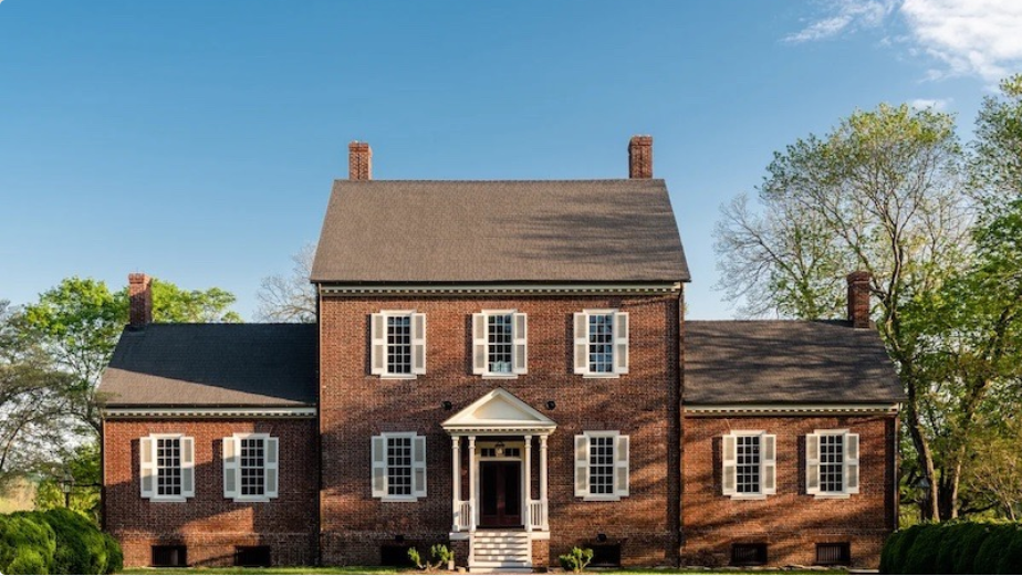
Located in Hillsborough, North Carolina, Ayr Mount strikes a startling figure in the historical town. The 1815 federal, masonry edifice was the first of its kind in this once bustling trade town, and thanks to the preservation efforts of our founder Richard Jenrette, remains well preserved with several original family objects on display.
CAHPT invites you to explore the site to learn about the design, construction, and maintenance of the estate as well as the lives of those, enslaved and free, who crafted and occupied the extravagant home and landscape from 1820 to 2018.
Visitors are welcome to explore the site select Thursdays, Fridays, and Saturdays April through November. Guided tours will be offered Thursday & Friday at 1:00pm and 2:30pm. Tour times for Saturday take place at 11:00am, 1:00pm, and 2:30pm. All guests must purchase tickets in advance.
Tickets can be purchased here: https://secure.classicalamericanhomes.org/nx/portal/neonevents/events#/events/
This is a potentially strenuous tour which includes climbing stairs, traversing uneven terrain, and enduring uncomfortable weather conditions and the presence of bugs or other wildlife. We regret that we cannot be responsible for those needing assistance and encourage all guests to dress accordingly. No food or beverages are allowed in the houses with the exception of a closed bottle of water.